Impetigo (impetigo contagiosa)
Exam relevance: in 2 of more than 50 board exam reports · rank 69
- Synonyms
- Borkenflechte, Grindflechte, Schleppe, bullöse Impetigo, Pyodermie, impetigo, school sores, impetigo contagiosa
- Specialty
- Dermatology · Bacterial infections
- Images
- Clinical 6
- In the app
- 4 flashcards
- Last updated
- 10/2026 · Dr. Pascal Bafteh
Contents
Images (6)
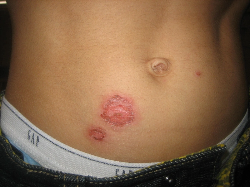
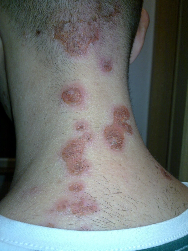
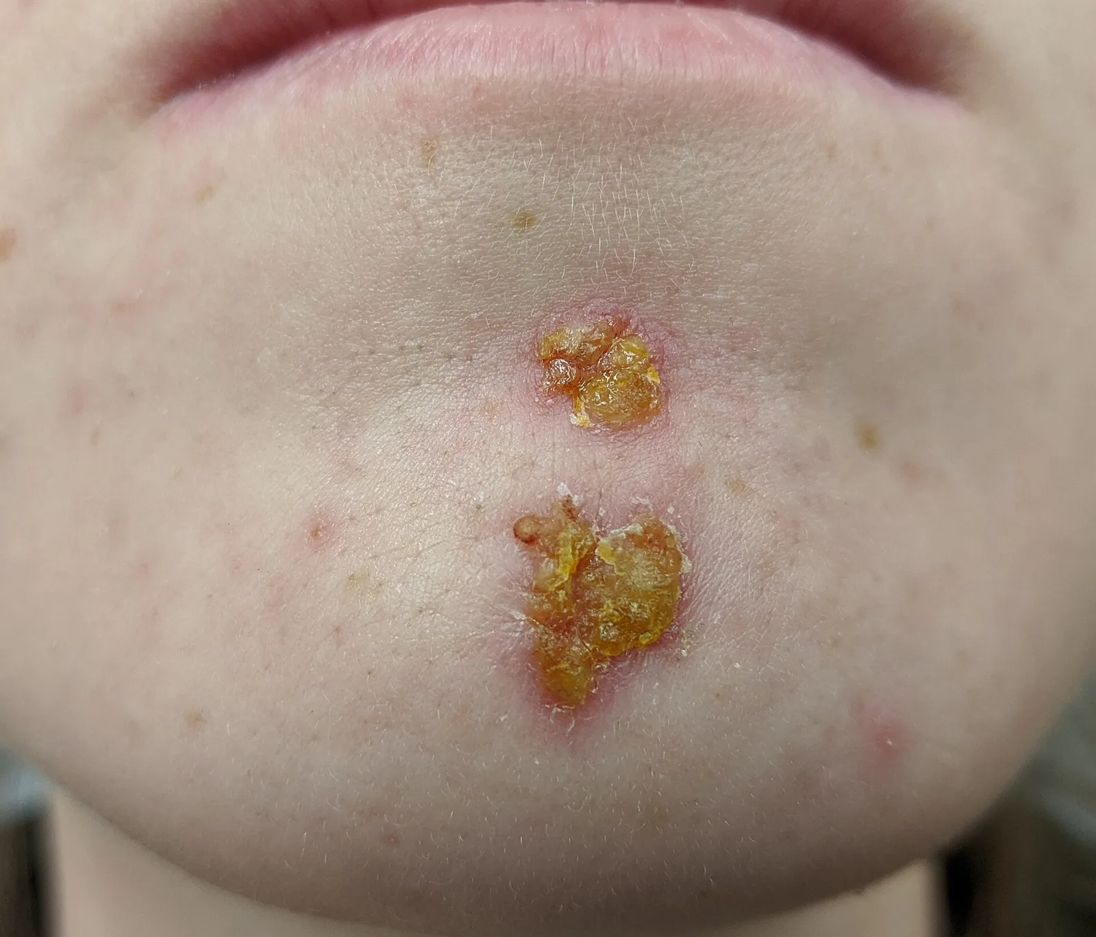
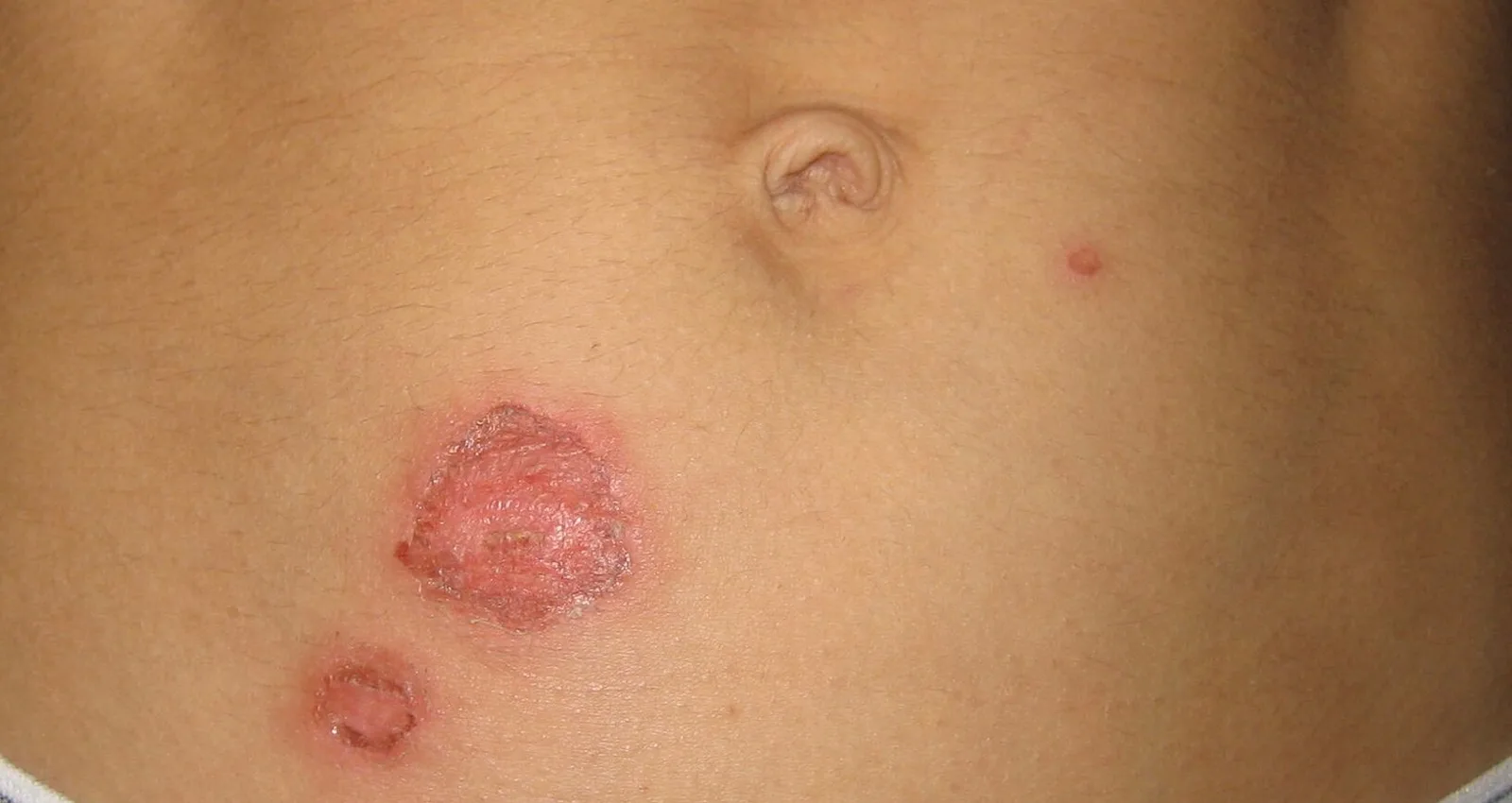
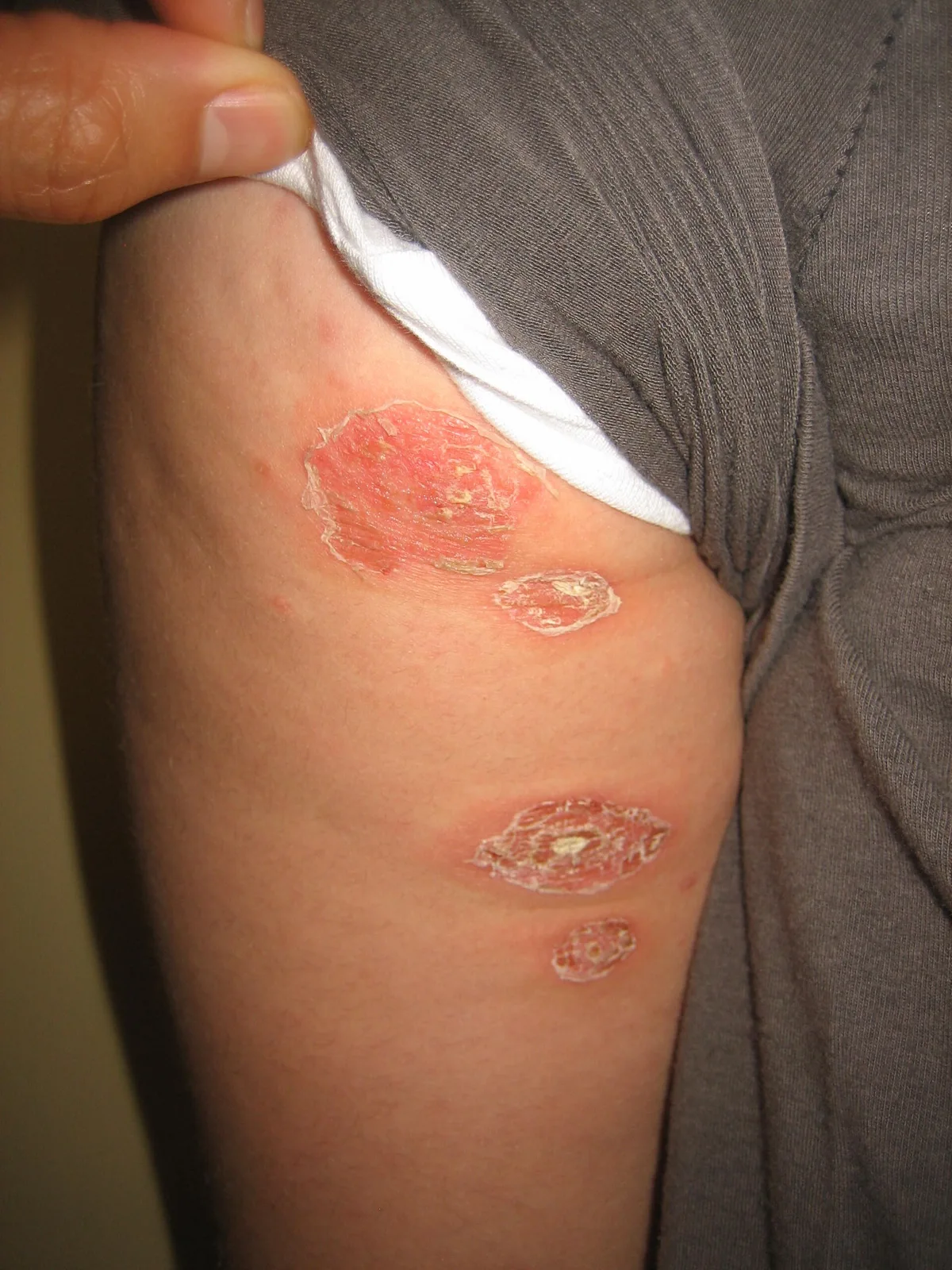
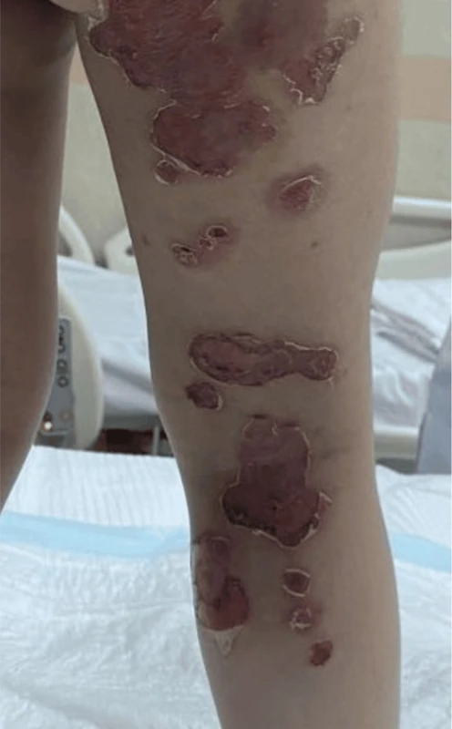
Definition
Impetigo (impetigo contagiosa) is a superficial, highly contagious bacterial infection of the epidermis caused by Staphylococcus aureus and/or Streptococcus pyogenes (group A beta-haemolytic streptococci).
Classification
- Non-bullous impetigo: the most common form, caused by S. aureus, streptococci or both.
- Bullous impetigo: caused by exfoliative toxins of S. aureus that cleave desmoglein 1 – the same mechanism as in staphylococcal scalded skin syndrome (SSSS), where, however, the toxins spread via the bloodstream.
- Impetiginisation: secondary infection of damaged skin, for example in atopic eczema, insect bites or scabies.
Occurrence & epidemiology
Impetigo is most common in young children but can occur at any age. It is usually transmitted through direct contact. Predisposing factors include skin conditions (atopic dermatitis, contact dermatitis, scabies, chickenpox), skin trauma (abrasions, insect bites, burns), immunosuppression, a warm humid climate, poor hygiene and crowded living conditions.
Clinical features
- Non-bullous: red macules with rapidly rupturing vesicles or pustules followed by characteristic honey-coloured crusts, mainly on the face (around the mouth and nose) and the limbs; little surrounding erythema; satellite lesions due to autoinoculation, "kissing lesions" where skin surfaces touch; systemic symptoms are usually absent, itch and regional lymphadenopathy may occur.
- Bullous: flaccid blisters with clear to cloudy content that rupture quickly, leaving erosions with a collarette of scale; face, trunk, limbs, buttocks and perineum; systemic symptoms (malaise, fever, lymphadenopathy) are more likely.
- Complications: deeper spread as ecthyma, lymphangitis, rarely post-streptococcal glomerulonephritis; SSSS with toxin-producing strains.
Histology
Non-bullous form (rarely biopsied): subcorneal collections of neutrophils, serum and parakeratosis; organisms are often hard to see on Gram stain. Bullous form: subcorneal blister with acantholytic cells in the upper epidermis, few neutrophils and sometimes Gram-positive cocci; a dermal inflammatory response in both.
Diagnosis
- Usually a spot diagnosis based on honey-coloured crusts or flaccid blisters.
- Bacterial swab with culture and sensitivity in recurrent or widespread disease or suspected MRSA; in recurrences a nasal swab for staphylococcal carriage; biopsy rarely (mainly in unclear bullous disease).
- Distinction from herpes simplex, pustular folliculitis, and pemphigus foliaceus and pemphigus vulgaris.
Differential diagnoses
 Erysipelas
 Erythrasma
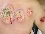 Pyoderma gangrenosum
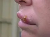 Herpes simplex (cold sores)
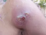 Furuncle and carbuncle
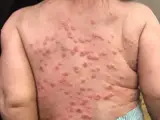 Eczema herpeticum
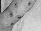 Ecthyma
Image credits for the thumbnails
- Erysipelas: Poupou l'quourouce, “Erysipel”, CC BY-SA 3.0, via Wikimedia Commons · CC BY-SA 3.0 · Source
- Erythrasma: unknown (Commons user), “Erythrasma”, CC BY-SA 4.0, via Wikimedia Commons · CC BY-SA 4.0 · Source
- Pyoderma gangrenosum: Monopol, “Pyoderma gangrenosum 01”, CC BY-SA 3.0, via Wikimedia Commons · CC BY-SA 3.0 · Source
- Herpes simplex (cold sores): unknown (Commons user), “Cold sore”, Public Domain, via Wikimedia Commons · Public Domain · Source
- Furuncle and carbuncle: Medicalpal, “Carbuncle”, CC BY-SA 4.0, via Wikimedia Commons · CC BY-SA 4.0 · Source
- Eczema herpeticum: Mohammad2018, CC BY-SA 4.0, via Wikimedia Commons, cropped · CC BY-SA 4.0 · Source
- Ecthyma: Norman Purvis Walker, An Introduction to Dermatology (3rd ed., 1905), public domain, via Wikimedia Commons – cropped · Public domain · Source
Treatment principles
- For localised disease, topical treatment with antiseptics, and if necessary topical antibiotics, after gentle removal of the crusts is usually sufficient.
- Extensive or bullous disease, systemic symptoms or spread warrant systemic antibiotic therapy effective against staphylococci and streptococci.
- Because of the high contagiousness, hygiene measures and the rules on attending childcare and school facilities must be observed.
Specific treatment regimens with drugs and doses are covered in the DermaFuchs app.
Keep learning in the app
References (selection)
Cross-references
Mentioned in
Keep learning
Keep learning with flashcards, exam questions and spot diagnoses in the DermaFuchs app – free, in your browser or as an app.
Related pages: Erysipelas · Erythrasma · Candida intertrigo
Note: This page is intended for medical education and does not replace diagnosis or treatment decisions in individual cases.