Necrotizing fasciitis
- Synonyms
- fleischfressende Bakterien, nekrotisierende Weichteilinfektion, necrotising fasciitis, necrotizing soft-tissue infection, flesh-eating disease, flesh-eating bacteria
- Specialty
- Dermatology · Bacterial infections
- Images
- Clinical 3 · Histology 1
- In the app
- 6 flashcards
- Last updated
- 10/2026 · Dr. Pascal Bafteh
Contents
Images (4)
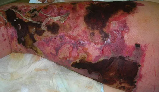
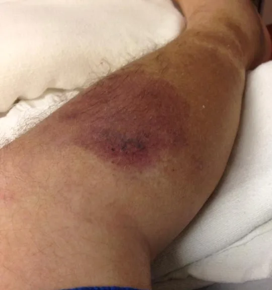
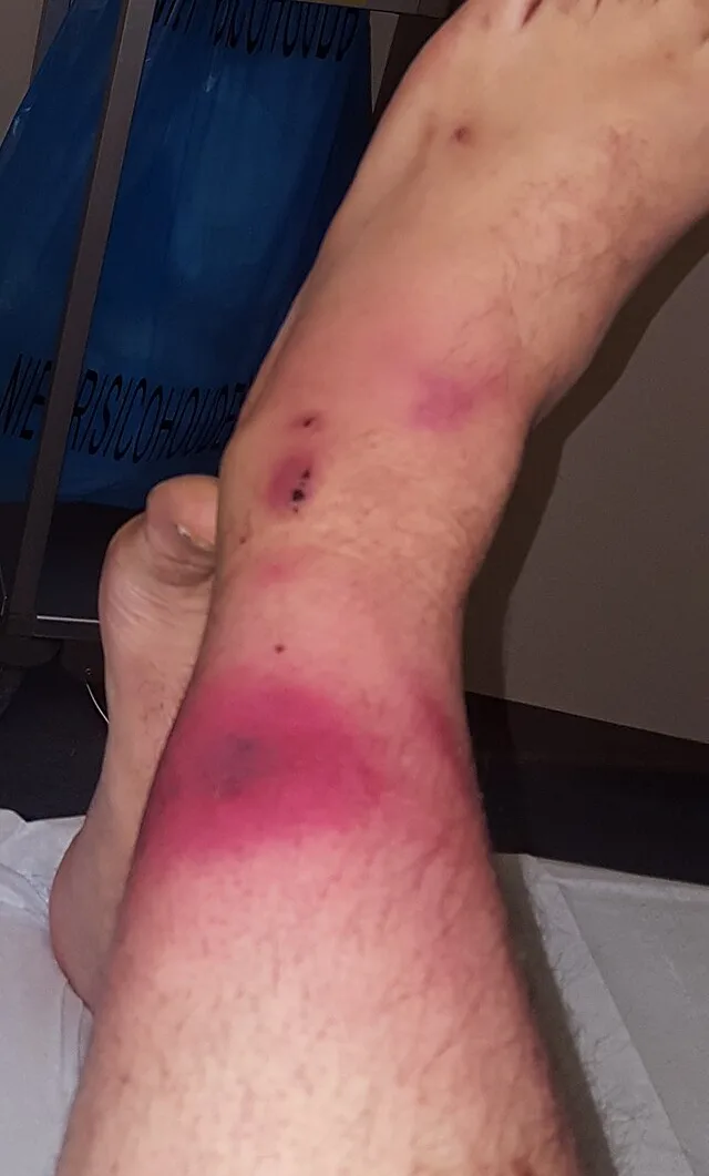
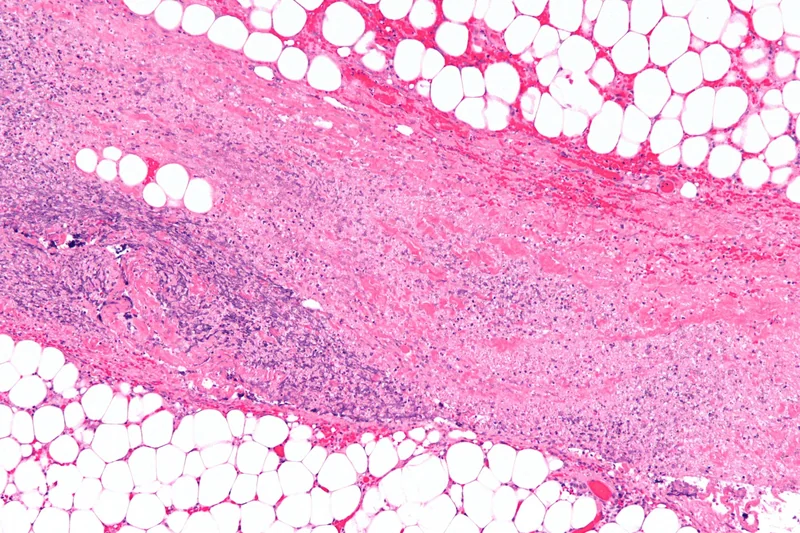 Histology
Definition
Necrotizing fasciitis is a life-threatening, rapidly progressive bacterial infection that destroys the subcutaneous tissue and fascia. Toxins and enzymes of the organisms cause thrombosis of small vessels and thus extensive tissue necrosis.
Classification
- Type I (polymicrobial): mixed aerobic and anaerobic flora (eg, Bacteroides, Escherichia coli), mainly on the trunk and perineum; common in older people and in diabetes mellitus.
- Type II (monomicrobial): mostly group A beta-haemolytic streptococci, second most commonly Staphylococcus aureus; any age, including previously healthy people; risk of toxic shock syndrome.
- Type III (in some classifications): gas gangrene due to clostridia, sometimes also marine organisms (Vibrio species, which other authors assign to type II). Type IV (in some classifications): fungi (Candida, zygomycetes) in immunocompromised people.
- Fournier gangrene: involvement of the perineum, genitals and perianal region.
Occurrence & epidemiology
Almost half of streptococcal cases occur in young, previously healthy people. The portal of entry is usually a skin injury, often a trivial one; in about 20% none is found. Risk factors include older age, diabetes mellitus, immunosuppression, obesity, chronic kidney disease, cancer, and alcohol and drug misuse; in children, type II can complicate chickenpox. In a meta-analysis, the mortality of necrotizing soft-tissue infections was 23.1%.
Clinical features
- The leading symptom is very severe pain out of proportion to the visible findings (with neuropathy, eg, diabetes, possibly mild); onset usually within 24 hours of an injury, often with fever and flu-like symptoms; most often on the lower leg.
- Initially redness, warmth and swelling resembling severe cellulitis; because the infection first spreads horizontally along the superficial fascia, the skin findings underestimate its extent.
- After 3–4 days, dusky discolouration, blisters with dark fluid, oedema, necrosis and crepitus from gas in the tissue; pain may subside once cutaneous nerves are destroyed.
- Rapid onset of tachycardia, hypotension and altered consciousness, progressing to septic or toxic shock.
Diagnosis
- Primarily a clinical diagnosis; the gold standard is exploration of the tissue: grey, dishwater-like exudate, friable superficial fascia, no pus and little bleeding. In the finger test, a finger passes through the subcutis without resistance via a small skin cut.
- Leucocytosis and raised CRP; blood cultures, tissue samples and Gram stain to identify the organism; X-ray, CT and MRI show the extent and gas in the tissue.
- LRINEC score (CRP, white cell count, haemoglobin, sodium, creatinine, glucose): a score of 6 or more makes necrotizing fasciitis likely (original study: positive predictive value 92%, negative predictive value 96%); because of poor sensitivity, a low score does not exclude it.
- Distinction: erysipelas involves the upper dermis and has a sharp, raised border, cellulitis the lower dermis and subcutis; necrotizing fasciitis extends to the fascia, with pain out of proportion to the findings and rapid deterioration.
Differential diagnoses
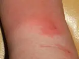 Erysipelas
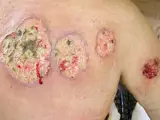 Pyoderma gangrenosum
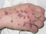 Purpura fulminans
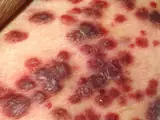 Kaposi's sarcoma
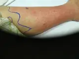 Cellulitis (phlegmon)
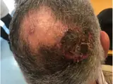 Cutaneous angiosarcoma
Image credits for the thumbnails
- Erysipelas: Poupou l'quourouce, “Erysipel”, CC BY-SA 3.0, via Wikimedia Commons · CC BY-SA 3.0 · Source
- Pyoderma gangrenosum: Monopol, “Pyoderma gangrenosum 01”, CC BY-SA 3.0, via Wikimedia Commons · CC BY-SA 3.0 · Source
- Purpura fulminans: Глей А.І., Шкурба А.В., Public domain, via Wikimedia Commons · Public domain · Source
- Kaposi's sarcoma: unknown author, “Kaposi's Sarcoma”, Public Domain, via Wikimedia Commons · Public Domain · Source
- Cellulitis (phlegmon): Stephen Ewen, “Left cellulitis of leg”, CC BY-SA 4.0, via Wikimedia Commons · CC BY-SA 4.0 · Source
- Cutaneous angiosarcoma: Messias H, Martins M, Zagalo C et al. (Cancer Rep (Hoboken) 2022), CC BY 4.0, via PubMed Central (PMC9575495) · CC BY 4.0 · Source
Treatment principles
- Necrotising fasciitis is a surgical emergency; immediate radical debridement of all necrotic tissue, usually with planned re-explorations, is crucial.
- At the same time, empirical intravenous broad-spectrum antibiotic therapy is started and adjusted once the pathogen is identified.
- Treatment takes place in intensive care; delaying surgery for further diagnostics considerably worsens the prognosis.
Specific treatment regimens with drugs and doses are covered in the DermaFuchs app.
Keep learning in the app
References (selection)
Cross-references
Mentioned in
Keep learning
Keep learning with flashcards, exam questions and spot diagnoses in the DermaFuchs app – free, in your browser or as an app.
Related pages: Abscess · Erysipelas · Pyoderma gangrenosum
Note: This page is intended for medical education and does not replace diagnosis or treatment decisions in individual cases.