Pyoderma gangrenosum
Exam relevance: in 7 of more than 50 board exam reports · rank 19
- Specialty
- Dermatology · Inflammatory skin diseases
- Images
- Clinical 4
- In the app
- 15 flashcards
- Last updated
- 10/2026 · Dr. Pascal Bafteh
Contents
Images (4)
Definition
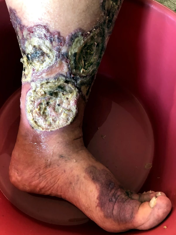
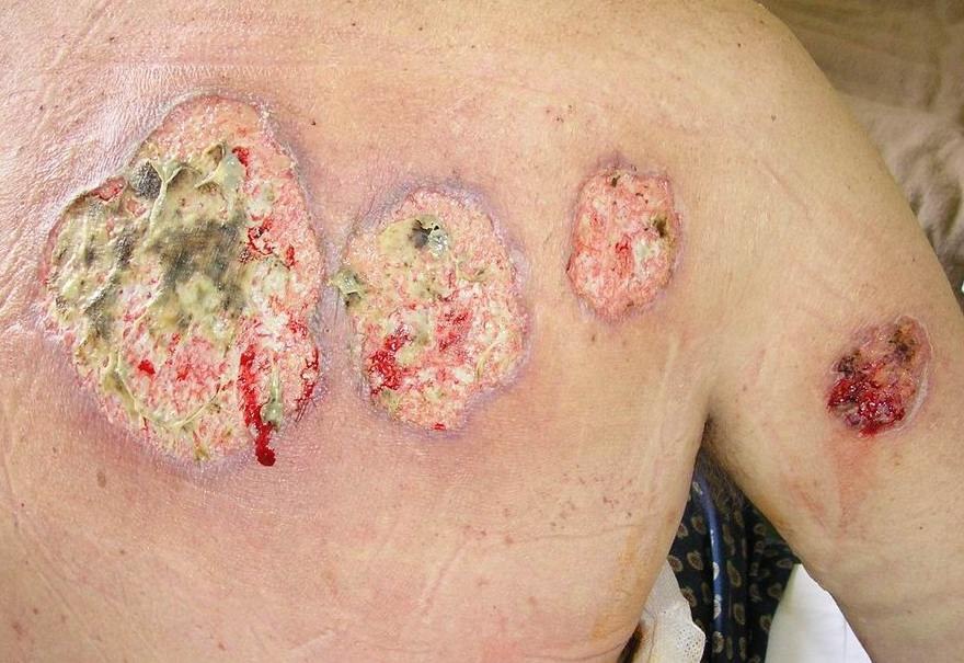
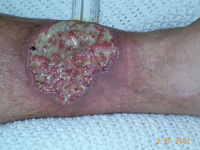
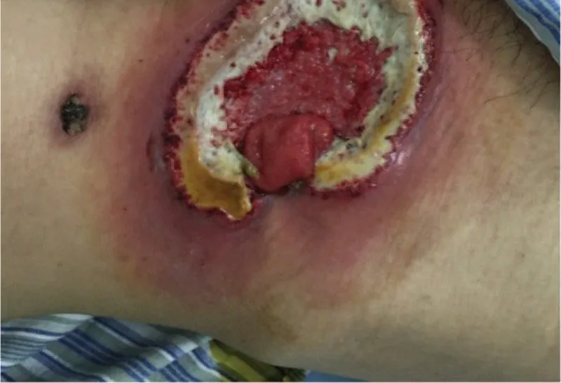
Pyoderma gangrenosum presents as a rapidly enlarging, very painful ulcer. It belongs to the autoinflammatory neutrophilic dermatoses; despite its historical name it is neither an infection nor gangrene. A full-thickness ulcer with blue-purple undermined borders and pathergy are characteristic.
- Women 20–50 years, neutrophilic dermatosis; livid eroded border + pathergy phenomenon + often peristomal (stoma) or post-traumatic.
- Diagnosis: Paracelsus Score / PARACELSUS Score — Points for: Major signs + Minor signs + Exclusion of other causes.
- Pathomechanism: neutrophilic inflammation via IL-1β / IL-36.
- Associations: PASH-/PAPASH-Syndrome (Pyoderma + Acne + Hidradenitis ± Pyogenic Arthritis), IBD, Hematological diseases.
Aetiopathogenesis
Pathogenesis & Clinical Features
- Pathogenesis: Pathogenesis involves IL-36 / IL-1β / IFN-γ (neutrophilic inflammation).
- Clinical Presentation: Atypical ulcer with a livid-erythematous border + peripheral pustules, often on the lower leg.
- Associations: IBD (ulcerative colitis, Crohn's disease) ~30%, RA, hematological neoplasms (MDS, AML, IgA-MGUS), PAPA/PASH spectrum.
Diagnosis
Diagnostics (Paracelsus Score)
- 3 Major Criteria (3 pts.): Rapidly progressive course, exclusion of other causes (PAD, DM ulcer, CVI, vasculitis), livid border.
- Minor and additional criteria (1–2 pts.): Improvement with immunosuppressants, irregular ulcer shape, extreme pain (VAS > 4/10), location at a site of trauma; suppurative inflammation on histology, undermined wound border, associated systemic disease.
- ≥ 10 points → probable PG.
- Spindle biopsy from the border for histology + exclusion of pathogens (caution: pathergy risk!).
Differential diagnoses
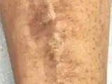 Diabetic dermopathy (shin spots)
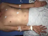 Secondary syphilis (lues II)
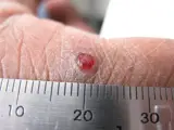 Pyogenic granuloma
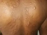 Lepromatous leprosy
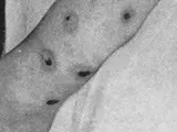 Ecthyma
Image credits for the thumbnails
- Diabetic dermopathy (shin spots): Abate, Aroucha, Nóbrega et al. (2025), “Cutaneous manifestations of diabetes mellitus: a narrative review”, CC BY 4.0, via Wikimedia Commons · CC BY 4.0 · Source
- Secondary syphilis (lues II): Ehlers S, Sergent S, Ashurst J (Clin Pract Cases Emerg Med 2020), CC BY 4.0, via PubMed Central (PMC7676796) · CC BY 4.0 · Source
- Pyogenic granuloma: kilbad, “Pyogenic granuloma on a finger-1”, CC BY 3.0, via Wikimedia Commons · CC BY 3.0 · Source
- Lepromatous leprosy: Shiyanbola AC, Emezie PC, Saheed O et al. (Cureus 2026), CC BY 4.0, via PubMed Central (PMC13294642) · CC BY 4.0 · Source
- Ecthyma: Norman Purvis Walker, An Introduction to Dermatology (3rd ed., 1905), public domain, via Wikimedia Commons – cropped · Public domain · Source
Treatment principles
- Because of pathergy, surgical debridement is avoided in the active phase; wound care is gentle and moist, with adequate analgesia.
- Localised lesions can be treated with topical or intralesional glucocorticoids or calcineurin inhibitors, and extensive disease systemically with glucocorticoids or calcineurin inhibitors.
- Biologics are used in refractory disease or with inflammatory bowel disease; associated underlying conditions are actively sought and treated.
Specific treatment regimens with drugs and doses are covered in the DermaFuchs app.
Keep learning in the app
References (selection)
Cross-references
Mentioned in
Keep learning
Keep learning with flashcards, exam questions and spot diagnoses in the DermaFuchs app – free, in your browser or as an app.
Related pages: Arterial (ischaemic) ulcer · Diabetic dermopathy (shin spots) · Secondary syphilis (lues II)
Note: This page is intended for medical education and does not replace diagnosis or treatment decisions in individual cases.