Inherited epidermolysis bullosa
Exam relevance: in 2 of more than 50 board exam reports · rank 69
- Synonyms
- hereditäre Epidermolysis bullosa, EB, Schmetterlingskinder, butterfly skin, epidermolysis bullosa
- Specialty
- Dermatology · Blistering diseases
- Images
- Clinical 6
- In the app
- 2 flashcards
- Last updated
- 10/2026 · Dr. Pascal Bafteh
Contents
Images (6)
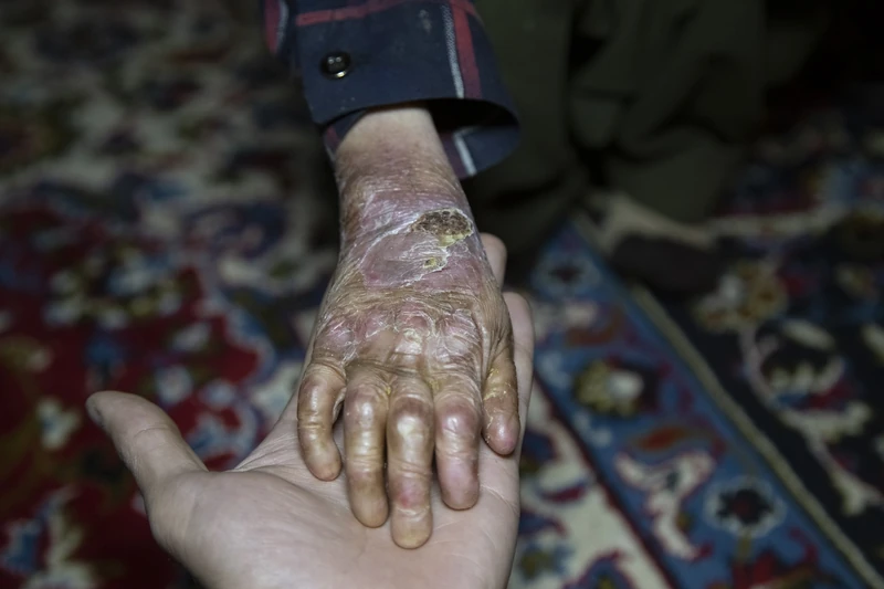
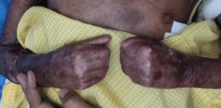
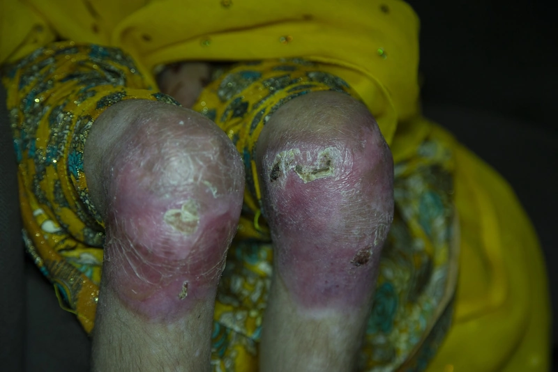
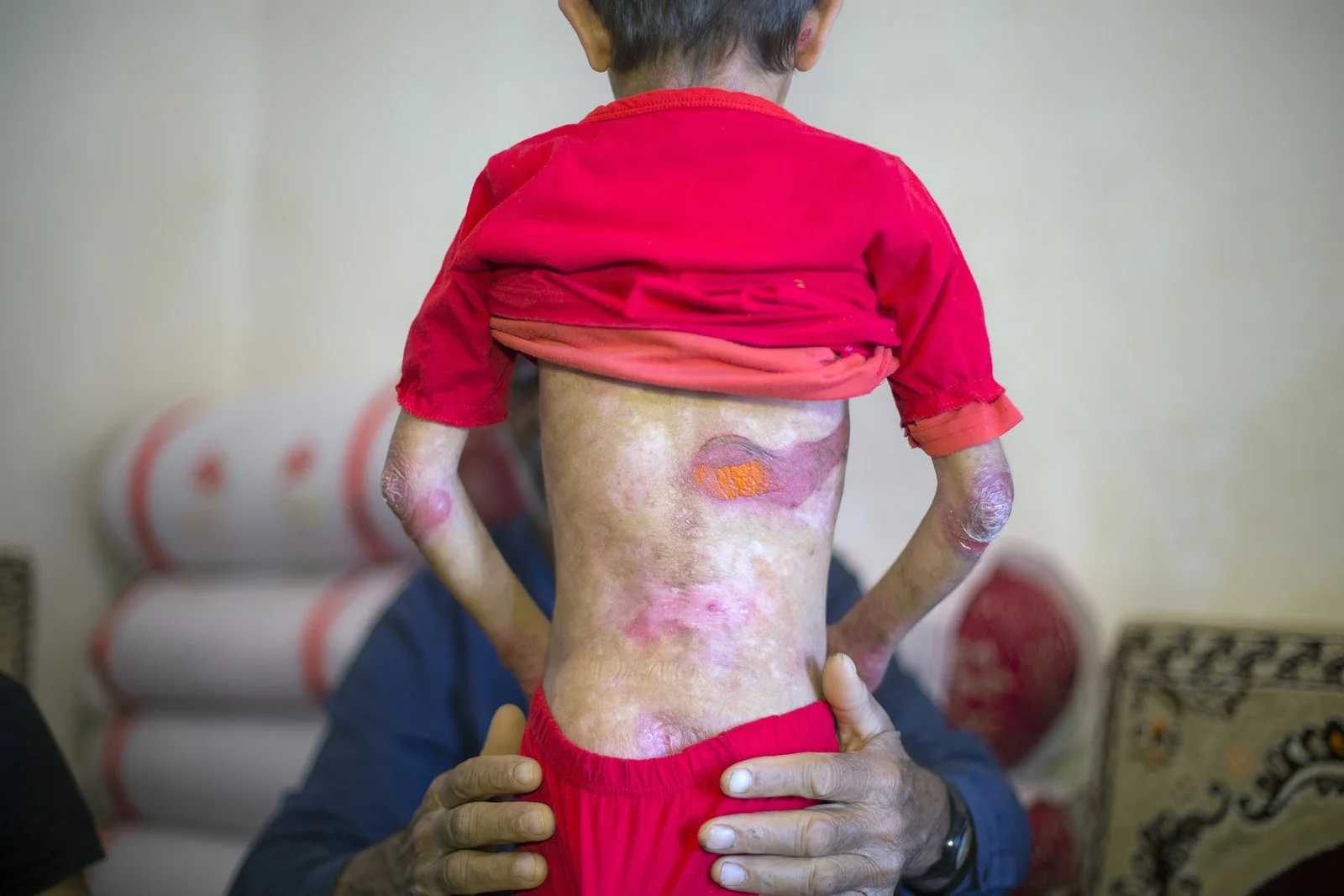
Definition
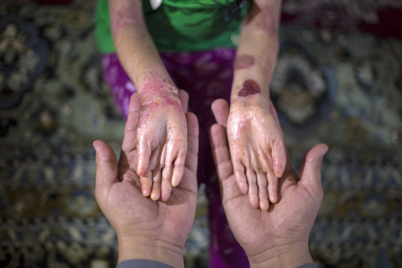
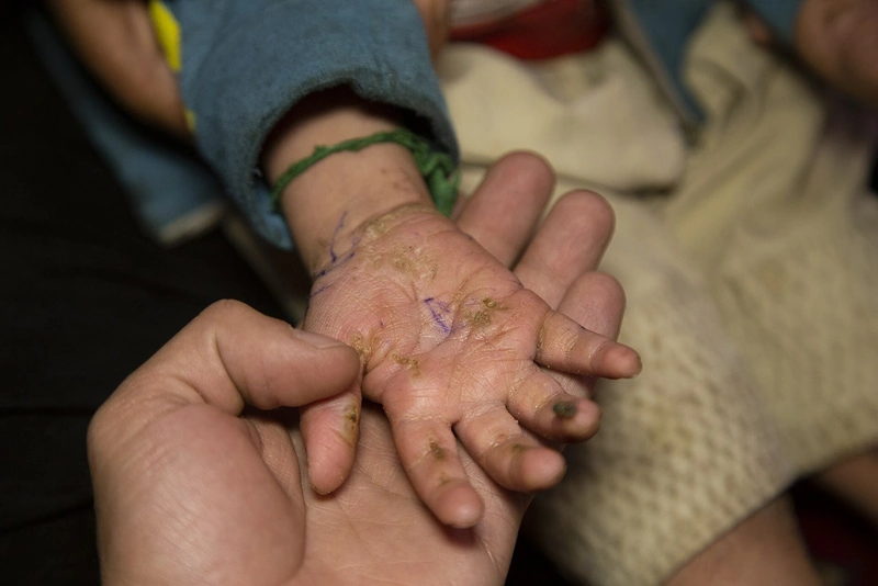
Inherited epidermolysis bullosa (EB) is a group of genetic disorders with skin fragility: even minor mechanical stress causes blisters, erosions and, depending on the type, scarring of the skin and mucous membranes. It is caused by variants in at least 16 genes encoding proteins that hold the layers of the skin together. It is distinct from acquired, autoimmune epidermolysis bullosa acquisita.
Classification
The 2020 international consensus distinguishes four main types by level of cleavage, with 34 subtypes in total:
- EB simplex (EBS): intraepidermal, within the basal keratinocytes; mainly keratin 5 and 14, less often plectin; mostly autosomal dominant; 14 subtypes.
- Junctional EB (JEB): within the lamina lucida of the basement membrane; mainly laminin 332 and collagen XVII, less often integrin alpha6beta4; nine subtypes.
- Dystrophic EB (DEB): below the lamina densa in the upper dermis; collagen VII (gene COL7A1), the main component of anchoring fibrils; dominant (DDEB) or recessive (RDEB) inheritance; 11 subtypes.
- Kindler EB: mixed level of cleavage (intraepidermal, junctional or below the lamina densa); kindlin-1 (gene FERMT1); autosomal recessive.
Other inherited skin fragility disorders in which blisters play little role have since been classified as separate groups.
Occurrence & epidemiology
In a large US survey, incidence and prevalence were 19.57 and 11.07 per million, respectively; EBS is the most common and JEB the rarest main type. The disease usually starts at or shortly after birth, although mild forms are sometimes recognised only later; both sexes are equally affected.
Clinical features
- EBS: blisters at sites of friction, especially on the hands and feet, which usually heal without scarring; localised to severe generalised forms.
- JEB: severe JEB (formerly Herlitz) shows generalised blisters and erosions from birth with mucosal involvement, exuberant granulation tissue around the nose and mouth, anaemia and susceptibility to infection, and is usually fatal in the first years of life; the intermediate form is milder and leaves subtle atrophy and hypopigmentation.
- DEB: from isolated nail dystrophy to generalised blistering with mutilating scarring. Severe RDEB (formerly Hallopeau-Siemens) shows pseudosyndactyly, joint contractures, chronic wounds, milia, oesophageal strictures, anaemia, developmental delay and a high risk of squamous cell carcinoma; dominant DEB is milder (eg, albopapuloid lesions, milia, atrophic scars).
- Kindler EB: blisters, photosensitivity, progressive poikiloderma and skin atrophy; gum erosions, involvement of the eyes, oesophagus, gut and genitourinary tract.
Diagnosis
- Biopsy of a newly induced blister: immunofluorescence mapping shows the level of cleavage and the absence or reduction of the affected protein; transmission electron microscopy assesses keratin filaments, hemidesmosomes, anchoring filaments and anchoring fibrils.
- Genetic testing (Sanger sequencing, gene panel, exome) establishes the subtype, mode of inheritance and causative variant; in localised DEB, the dominant and recessive forms can only be distinguished genetically.
Differential diagnoses
 Pemphigus vulgaris
 Linear IgA dermatosis
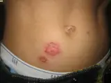 Impetigo (impetigo contagiosa)
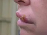 Herpes simplex (cold sores)
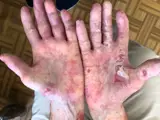 Epidermolysis bullosa acquisita
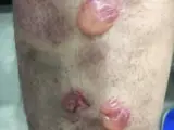 Bullous pemphigoid
Image credits for the thumbnails
- Pemphigus vulgaris: Mohammad2018, CC BY-SA 4.0, via Wikimedia Commons, cropped · CC BY-SA 4.0 · Source
- Linear IgA dermatosis: Mohammad2018, “IgA bullous dermatosis”, CC BY-SA 4.0, via Wikimedia Commons · CC BY-SA 4.0 · Source
- Impetigo (impetigo contagiosa): Littlekidsdoc, “Bullous impetigo”, CC BY-SA 4.0, via Wikimedia Commons · CC BY-SA 4.0 · Source
- Herpes simplex (cold sores): unknown (Commons user), “Cold sore”, Public Domain, via Wikimedia Commons · Public Domain · Source
- Epidermolysis bullosa acquisita: Szymański, Kowalewski, Pietrzyk et al. (2023), “Case Report: Biological treatment of epidermolysis bullosa acquisita: report on four cases and literature review”, CC BY 4.0, via Wikimedia Commons · CC BY 4.0 · Source
- Bullous pemphigoid: Mohammad2018, “Bullous pemphigoid new image”, CC BY-SA 4.0, via Wikimedia Commons · CC BY-SA 4.0 · Source
Treatment principles
- A causal therapy is not yet widely available; management centres on specialised wound care with non-adherent dressings and avoidance of mechanical stress.
- Supportive measures include adequate nutrition to meet the high calorie requirement, pain management and, where necessary, surgical release of finger contractures.
- In dystrophic forms, regular surveillance for squamous cell carcinoma is necessary; care is provided in specialised centres.
Specific treatment regimens with drugs and doses are covered in the DermaFuchs app.
Keep learning in the app
References (selection)
Cross-references
Keep learning: test yourself
One question from the DermaFuchs app on Inherited epidermolysis bullosa – tap an answer.
Question 1 of 1
Which proteins are involved in EB simplex affecting basal keratinocytes?
Answer
Correct: Keratin 5 / 14, Plectin. EB simplex involves defects in Keratin 5/14 and Plectin, affecting the structural integrity of basal keratinocytes.
More questions like these – with explanations and spaced repetition – in the app.
Note: This page is intended for medical education and does not replace diagnosis or treatment decisions in individual cases.