Cellulitis (phlegmon)
- Synonyms
- Zellulitis, Cellulitis, Weichteilphlegmone, Unterschenkelphlegmone, phlegmon, soft tissue infection
- Specialty
- Dermatology · Bacterial infections
- Images
- Clinical 1
- In the app
- 4 flashcards
- Last updated
- 10/2026 · Dr. Pascal Bafteh
Contents
Images (1)
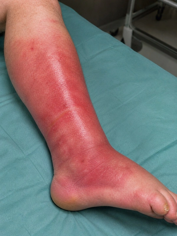 AI illustration
Definition
Cellulitis (phlegmon) is an acute, diffusely spreading bacterial infection of the deep dermis and subcutaneous tissue.
Distinction from erysipelas: erysipelas is a more superficial infection (upper dermis including the lymphatics) with shiny, raised, sharply demarcated redness; cellulitis lies deeper and usually has indistinct borders. In some regions, notably Europe, the terms erysipelas and cellulitis are also used synonymously.
Aetiopathogenesis
The most common organisms are group A beta-haemolytic streptococci (Streptococcus pyogenes, about two thirds) and Staphylococcus aureus (about one third). Streptococci spread diffusely and rapidly by means of enzymes such as streptokinase, DNase and hyaluronidase; staphylococcal infection is more localised and often arises from open wounds or abscesses. Portals of entry are breaks in the skin, for example from tinea pedis, onychomycosis, venous leg ulcers, pressure injuries or scratched eczema; further risk factors are previous episodes, lymphoedema, immunodeficiency, diabetes and obesity.
Clinical features
- Mostly on the lower limb and unilateral; bilateral involvement suggests another cause.
- Warm, red, oedematous, painful skin, often with peau d'orange; the borders are usually indistinct – unlike the sharply demarcated border of erysipelas.
- Petechiae are common; blisters, erosions and necrosis can occur. Usually non-purulent; the purulent form is accompanied by pustules, furuncles or abscesses.
- Fever and chills may precede the skin findings by hours; lymphangitis and regional lymphadenopathy are common.
- Warning signs of complicated or necrotising infection: pain out of proportion to the findings, cutaneous haemorrhage, bullae, skin sloughing, skin anaesthesia, rapid progression, tissue gas, signs of systemic toxicity.
Diagnosis
- Clinical diagnosis from history and examination; blood tests often show leucocytosis and raised CRP.
- Blood and tissue cultures are not routine but are useful in immunosuppression or with systemic signs of infection.
- Imaging: ultrasound to distinguish an abscess, MRI if necrotising soft tissue infection is suspected, duplex ultrasound if deep vein thrombosis is suspected.
- Distinction from, among others, stasis dermatitis (usually bilateral) and contact dermatitis (itch, limited to the contact site).
Differential diagnoses
Keep learning in the app
References (selection)
Cross-references
Note: This page is intended for medical education and does not replace diagnosis or treatment decisions in individual cases.