Cutaneous myiasis
- Synonyms
- myiasis, botfly, maggot infestation
- Specialty
- Dermatology · Parasitic skin diseases
- Images
- Clinical 3
- In the app
- 1 flashcard
- Last updated
- 10/2026 · Dr. Pascal Bafteh
Contents
Images (3)
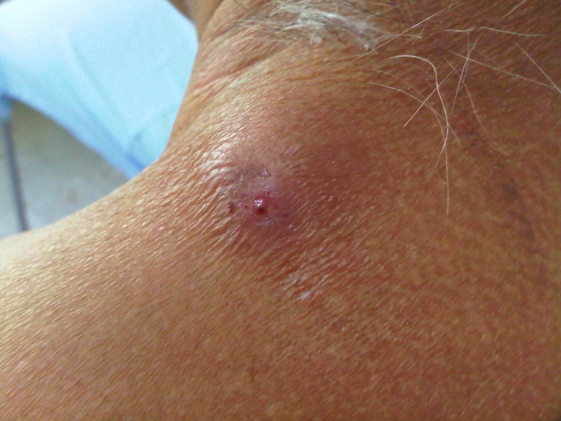
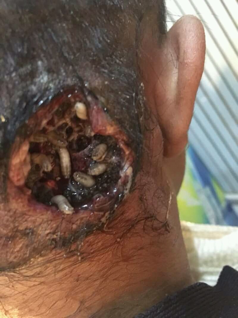
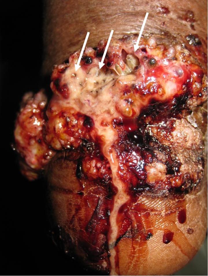
Definition
Myiasis is the infestation of skin or body cavities by fly larvae. Furuncular, migratory and wound myiasis are distinguished. Common causes of furuncular myiasis are Dermatobia hominis (Central and South America) and Cordylobia anthropophaga (tropical Africa); the travel history is important for the diagnosis. In wound myiasis fly larvae infest open wounds.
- Causative agent: Dermatobia + Cordylobia (Human botfly and Tumbu fly).
- Transmission: Dermatobia lays its eggs on carrier insects (mostly mosquitoes) or foliage, Cordylobia on objects contaminated with urine or faeces such as laundry or soil.
- Clinical presentation: painful, erythematous papule/nodule with central breathing hole — foreign body sensation, often movement of the larva palpable.
- Diagnosis: clinical + visualization of the larva (ultrasound helpful).
Differential diagnoses
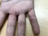
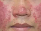
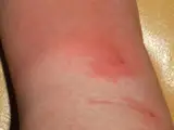
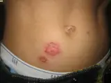
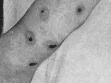
Image credits for the thumbnails
- Furuncle and carbuncle: Medicalpal, “Carbuncle”, CC BY-SA 4.0, via Wikimedia Commons · CC BY-SA 4.0 · Source
- Scabies: Gzzz, “Scabies hand and fingers 1”, CC BY-SA 4.0, via Wikimedia Commons · CC BY-SA 4.0 · Source
- Demodicosis: Ran Yuping et al., “Demodicosis”, CC BY-SA 3.0, via Wikimedia Commons · CC BY-SA 3.0 · Source
- Erysipelas: Poupou l'quourouce, “Erysipel”, CC BY-SA 3.0, via Wikimedia Commons · CC BY-SA 3.0 · Source
- Impetigo (impetigo contagiosa): Littlekidsdoc, “Bullous impetigo”, CC BY-SA 4.0, via Wikimedia Commons · CC BY-SA 4.0 · Source
- Ecthyma: Norman Purvis Walker, An Introduction to Dermatology (3rd ed., 1905), public domain, via Wikimedia Commons – cropped · Public domain · Source
Treatment principles
- Complete removal of the larvae is crucial, for example after occluding the breathing pore, by extraction or surgical incision.
- Wound care follows; secondary infections are treated with antibiotics, and tetanus immunisation status should be checked.
- In wound myiasis, thorough debridement and treatment of predisposing factors such as inadequate wound care are important.
Specific treatment regimens with drugs and doses are covered in the DermaFuchs app.
Keep learning in the app
References (selection)
Cross-references
Mentioned in
Keep learning: test yourself
One question from the DermaFuchs app on Cutaneous myiasis – tap an answer.
Question 1 of 1
Which causative agents are responsible for human myiasis, specifically the human botfly and Tumbu fly?
Answer
Correct: Dermatobia + Cordylobia. Dermatobia (human botfly) and Cordylobia (Tumbu fly) are the primary causative agents of cutaneous myiasis.
More questions like these – with explanations and spaced repetition – in the app.
Note: This page is intended for medical education and does not replace diagnosis or treatment decisions in individual cases.