Erythrasma
- Synonyms
- Corynebacterium minutissimum
- Specialty
- Dermatology · Bacterial infections
- Images
- Clinical 1
- In the app
- 6 flashcards
- Last updated
- 10/2026 · Dr. Pascal Bafteh
Contents
Images (1)
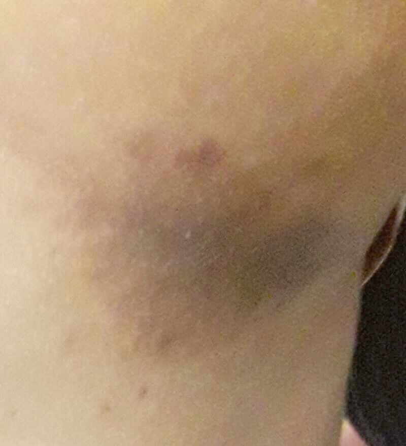
Definition
Typical are brownish-red, finely scaling plaques intertriginous in axilla / groin / submammary / interdigital.
Pathogen: Corynebacterium minutissimum (gram-positive rod, porphyrin-producing).
Diagnostics: Characteristic is coral-red fluorescence under Wood's light due to Coproporphyrin III.
DD: Tinea cruris (KOH+, peripheral scaling), Candidiasis intertriginosa, inverse psoriasis, Hailey-Hailey disease.
Occurrence & epidemiology
Erythrasma is a common superficial bacterial infection of the skin folds (axillae, groins, toe webs) caused by Corynebacterium minutissimum. Predisposing factors are warm climate, excessive sweating, obesity, diabetes mellitus (widespread forms), older age and immunocompromise. Interdigital (3rd–5th toe webs), intertriginous and generalised/disciform (trunk) forms are distinguished.
Clinical features
- Sharply demarcated, pink to brown patches with fine scaling and superficial fissures.
- Mild itch may be present; may coexist with or be confused with tinea or candidal intertrigo.
Differential diagnoses
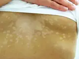
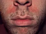
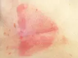
Image credits for the thumbnails
- Pityriasis versicolor (tinea versicolor): Sarahrosenau on Flickr.com, “Tinea versicolor1”, CC BY-SA 2.0, via Wikimedia Commons · CC BY-SA 2.0 · Source
- Seborrhoeic dermatitis: Roymishali, “Seborrhoeic dermatitis highres”, CC BY-SA 3.0, via Wikimedia Commons · CC BY-SA 3.0 · Source
- Intertrigo: Pera F, Suman C, Cosma M, Mazza S, Brunani A, Cancello R (J Cosmet Dermatol 2025), CC BY 4.0, via PubMed Central (PMC12042251) – panel B “before” (cropped) · CC BY 4.0 · Source
Treatment principles
- Treatment is topical, for example with azole antifungals, which are also active against corynebacteria, or with topical antibiotics.
- Extensive or recurrent disease may require systemic antibiotic therapy.
- To prevent relapse, predisposing factors such as warmth and moisture, sweating in skin folds and obesity are addressed; antiseptic washes can be used as an adjunct.
Specific treatment regimens with drugs and doses are covered in the DermaFuchs app.
Keep learning in the app
References (selection)
Cross-references
Mentioned in
Keep learning: test yourself
One question from the DermaFuchs app on Erythrasma – tap an answer.
Question 1 of 1
What characteristic fluorescence color does Erythrasma show under Wood's light examination?
Answer
Correct: coral red. Corynebacterium minutissimum produces coproporphyrin III, which fluoresces coral red under ultraviolet Wood's light.
More questions like these – with explanations and spaced repetition – in the app.
Note: This page is intended for medical education and does not replace diagnosis or treatment decisions in individual cases.