Epidermolysis bullosa simplex
- Synonyms
- EBS, Schmetterlingskinder, butterfly children, epidermolysis bullosa
- Specialty
- Dermatology · Hereditary skin diseases
- Images
- Clinical 2
- In the app
- 7 flashcards
- Last updated
- 10/2026 · Dr. Pascal Bafteh
Contents
Images (2)
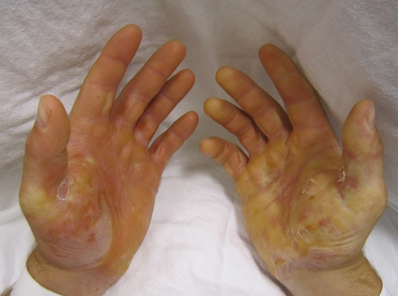
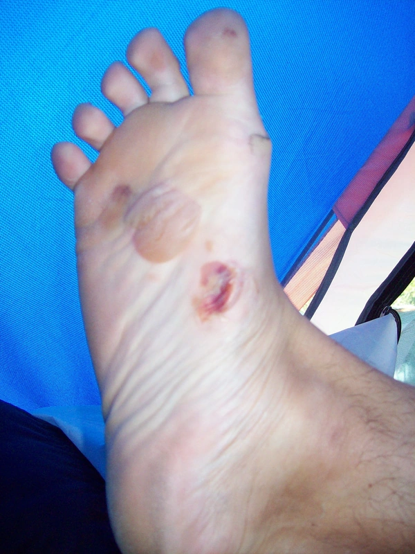
Definition
Epidermolysis bullosa simplex (EBS) is a major type of inherited epidermolysis bullosa. Skin cleavage occurs within the epidermis, inside the basal keratinocytes; even minor mechanical stress causes blisters and erosions.
Occurrence & epidemiology
In a large US cohort, EBS had the highest incidence and prevalence of all EB types (7.87 and 6.0 per million, respectively); the localised form is the most common. Because mild forms are easily missed, the prevalence is probably higher. Most forms are inherited in an autosomal dominant manner, although recessive inheritance is more common in some regions; both sexes are equally affected.
Aetiopathogenesis
Keratins 5 and 14 form the intermediate filament cytoskeleton of basal keratinocytes. Variants in the KRT5 or KRT14 genes – mostly missense variants with autosomal dominant inheritance – disrupt its assembly and cause the three main subtypes (60–70% of EBS cases); variants at the ends of the helical rod domains are often associated with severe forms. Variants in the plectin gene (PLEC) account for up to 8% of cases. Overall, at least seven genes and 14 clinical subtypes have been described.
Clinical features
- Localised EBS (formerly Weber-Cockayne): blisters on the palms and soles or at sites of repeated friction, usually from crawling and walking age, worse in warm months; in adulthood sometimes focal palmoplantar keratoderma; the most common complication is infection of blisters on the feet.
- Intermediate EBS (formerly Koebner): often generalised blisters from birth, predominantly on the hands and feet, without herpetiform clustering and without extracutaneous involvement; worse with heat.
- Severe EBS (formerly Dowling-Meara): large, generalised, partly herpetiform and haemorrhagic blisters, oral mucosal involvement, progressive palmoplantar keratoderma and nail dystrophy; blistering improves with age.
- Blisters usually heal without scarring; milia and hypo- or hyperpigmentation may occur.
- Rare PLEC-related forms: EBS with muscular dystrophy and severe EBS with pyloric atresia.
Diagnosis
- With a typical picture and an informative family tree with dominant inheritance, a clinical diagnosis is often possible.
- Immunofluorescence mapping and transmission electron microscopy of a newly induced blister determine the level of cleavage.
- Genetic testing establishes the subtype, mode of inheritance and causative variant.
Differential diagnoses
 Epidermolysis bullosa acquisita
 Porphyria cutanea tarda
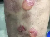 Bullous pemphigoid
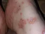 Linear IgA dermatosis
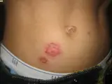 Impetigo (impetigo contagiosa)
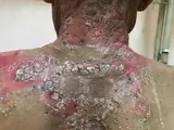 Pemphigus vulgaris
Image credits for the thumbnails
- Epidermolysis bullosa acquisita: Szymański, Kowalewski, Pietrzyk et al. (2023), “Case Report: Biological treatment of epidermolysis bullosa acquisita: report on four cases and literature review”, CC BY 4.0, via Wikimedia Commons · CC BY 4.0 · Source
- Porphyria cutanea tarda: Nicolás Hermosilla B.; Gonzalo de Toro; Montserrat Molgó, “Blister in porphyria cutanea tarda”, CC BY 4.0, via Wikimedia Commons · CC BY 4.0 · Source
- Bullous pemphigoid: Mohammad2018, “Bullous pemphigoid new image”, CC BY-SA 4.0, via Wikimedia Commons · CC BY-SA 4.0 · Source
- Linear IgA dermatosis: Mohammad2018, “IgA bullous dermatosis”, CC BY-SA 4.0, via Wikimedia Commons · CC BY-SA 4.0 · Source
- Impetigo (impetigo contagiosa): Littlekidsdoc, “Bullous impetigo”, CC BY-SA 4.0, via Wikimedia Commons · CC BY-SA 4.0 · Source
- Pemphigus vulgaris: Mohammad2018, CC BY-SA 4.0, via Wikimedia Commons, cropped · CC BY-SA 4.0 · Source
Treatment principles
- There is no causal treatment; management is symptomatic.
- The main measures are avoidance of friction and heat, which promote blistering, well-fitting footwear and gentle wound care, in which tense blisters are lanced under sterile conditions.
- Genetic counselling is offered.
Specific treatment regimens with drugs and doses are covered in the DermaFuchs app.
Keep learning in the app
References (selection)
Cross-references
Keep learning
Keep learning with flashcards, exam questions and spot diagnoses in the DermaFuchs app – free, in your browser or as an app.
Related pages: Epidermolysis bullosa acquisita · Porphyria cutanea tarda · Bullous pemphigoid
Note: This page is intended for medical education and does not replace diagnosis or treatment decisions in individual cases.