Lepromatous leprosy
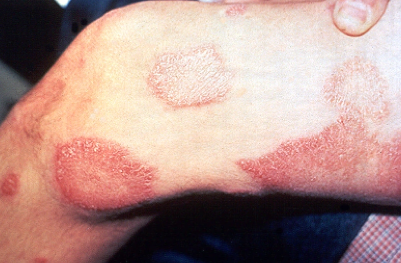
Pathogen & Pathogenesis
- The pathogen is M. leprae, an acid-fast bacterium, obligate intracellular with a generation time of ~14 days — only in animal models (armadillo).
- Transmission: aerogenic droplets with low contagiousness and an incubation period of 5–20 years.
- Tropism: cooler body regions (skin, peripheral nerves, nasal mucosa) due to the pathogen's heat sensitivity.
Classification (Ridley-Jopling, Immunomodulated Spectrum Form)
| Form & Immune status | Clinical features & Localization |
|---|---|
| TT — Tuberculoid · strong Th1 | 1–3 sharply demarcated, hypopigmented/erythematous plaques with anesthesia and anhidrosis, central atrophy. Few areas. |
| BT — Borderline Tuberculoid · Th1 dominant | 3–10 plaques, moderate extent. |
| BB — Borderline · mixed Th1/Th2 | Many lesions, unstable course, generalized. |
| BL — Borderline Lepromatous · Th2 dominant | Many lesions, generalized. |
| LL — Lepromatous · weak Th2 + anergy | Diffuse infiltrative thickening, facies leontina, madarosis (lateral eyebrows), saddle nose, numerous nodules, high bacterial load, generalized symmetrical. |
Key Clinical Features
Tuberculoid Leprosy (TT)
- Hypopigmented or erythematous, sharply demarcated, anhidrotic plaques with central atrophy.
- Pathognomonic is loss of sensation with impaired temperature/pressure/pain — Early sign: temperature discrimination ↓.
- Nearby peripheral nerves thickened (e.g., ulnar nerve, great auricular nerve, common peroneal nerve) palpable.
Lepromatous Leprosy (LL)
- Diffuse skin infiltration with livid nodules, facies leontina, madarosis, saddle nose (nasal mucosa involvement with cartilage destruction), testicular atrophy (M. leprae involvement).
- Peripheral neuropathy with claw hand, foot drop.
- Complications: Glaucoma, keratitis, iridocyclitis (eye involvement), bone destruction (acro-osteolysis), secondary infections.
Diagnostics
- Clinical presentation + sensation testing (cotton, needle, temperature).
- Skin biopsy (skin lesion + nerve biopsy) with:
- Ziehl-Neelsen staining (acid-fast bacilli detection in lepromatous form)
- Fite-Faraco staining more sensitive for M. leprae
- Quantification is performed via Bacillary Index on mucosal/skin smear with a scale 0–6+ logarithmic for classification/therapy monitoring.
- PCR (M. leprae DNA) as a sensitive pathogen detection method in paucibacillary forms.
- Lepromin skin test (historical) — shows Th1 reactivity, not for diagnosis but for classification.
- Negative control: sensory examination compared to the contralateral skin area.
Epidemiology & Reporting Obligation
- WHO elimination as a "Public Health Problem" achieved in 2000 (< 1 case / 10,000), but endemic in India, Brazil, Indonesia, Nepal, Africa (~ 200,000 new cases per year worldwide).
- In DE, there is IfSG §7 by name with reporting to health authority.
- DTG/RKI recommendation: Travel history for any unclear hypopigmented skin finding with loss of sensation.
Differential diagnoses
- Mycosis fungoides
- Tinea corporis
- Cutaneous leishmaniasis
- Lupus erythematosus gesicht
- Cutaneous lymphoma
- Erythema nodosum
- Erythema migrans (Lyme disease)
- Cutaneous sarcoidosis
Practise Lepromatous leprosy in the app
Flashcards with spaced repetition, exam questions and spot-the-diagnosis on this topic – in DermaFuchs, free of charge.
In the DermaFuchs app: 2 flashcards · 5 clinical images
Open in browser Download on the App StoreSources (selection)
More bacterial skin infections
Note: This page is intended for medical education and does not replace diagnosis or treatment decisions in individual cases. Treatment and follow-up content is available in the app.