Herpes zoster
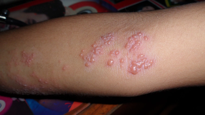
![Herpes zoster: unilateral dermatomal groups of vesicles on the flank (day 3) [Bild 180° gedreht 16.09.2026 — Original stand auf dem Kopf]](/assets/img/wissen/herpes-zoster-2.webp)
Definition and Pathogenesis
Herpes zoster is caused by the reactivation of the varicella-zoster virus, a DNA virus from the family Herpesviridae. After a primary infection (varicella), the virus persists latently in the spinal ganglia and can be reactivated when cellular immunity wanes.
Epidemiology and Risk Factors
The disease occurs more frequently in:
- older age (>60 yrs)
- immunosuppression
- HIV infection
Clinical Presentation
Typical is a unilateral dermatomal rash with grouped vesicles on an erythematous base. Often, burning pain precedes the skin changes by several days.
Most commonly affected dermatomes are in the area of:
- thorax (approx. 50–70 % of cases, especially Th3–L3)
- trigeminal nerve (V1 > V2 > V3) — high ocular complication rate with V1 involvement
Special Forms
Zoster ophthalmicus results from involvement of the first trigeminal branch and can lead to keratitis. The appearance of lesions on the tip of the nose is called Hutchinson sign (involvement of the nasociliary nerve) and indicates ocular involvement.
- In case of ocular involvement (zoster ophthalmicus, V1) MANDATORY ophthalmology consult — risk of keratitis, iridocyclitis, acute retinal necrosis (ARN), vision loss.
- In case of ear involvement (zoster oticus / Ramsay-Hunt syndrome) MANDATORY ENT consult — risk of peripheral facial nerve palsy, sensorineural hearing loss, vestibulopathy/vertigo.
Complications
The most common complication is post-zoster neuralgia, classically defined as persistent pain lasting longer than 4 weeks after resolution of the skin lesions; the German S2k guideline and the IASP set the cut-off at > 3 months after rash onset (30–90 days = subacute zoster neuralgia). Further possible complications include bacterial superinfection of the lesions and — depending on the affected nerve — peripheral facial nerve palsy, sensorineural hearing loss and iridocyclitis/uveitis.
- Zoster meningoencephalitis (rare, ~0.1–0.5 %): usually 1–2 weeks after rash onset. Clinical: headache, fever, meningismus, confusion. Diagnosis: VZV PCR in CSF. Therapy: aciclovir i.v. 10 mg/kg body weight every 8 h for 14–21 days. Increased risk: immunosuppressed, multidermatomal zoster, trigeminal involvement.
Differential diagnoses
- Herpes simplex labialis
- Phlegmon
- Varicella (chickenpox)
- Eczema herpeticum
- Impetigo
- Phytophotodermatitis
- Erysipelas
Practise Herpes zoster in the app
Flashcards with spaced repetition, exam questions and spot-the-diagnosis on this topic – in DermaFuchs, free of charge.
In the DermaFuchs app: 7 flashcards · 14 clinical images
Open in browser Download on the App StoreSources (selection)
More viral skin infections
Note: This page is intended for medical education and does not replace diagnosis or treatment decisions in individual cases. Treatment and follow-up content is available in the app.