Linear IgA dermatosis
- Synonyms
- linear IgA disease, chronic bullous disease of childhood
- Specialty
- Dermatology · Blistering diseases
- Images
- Clinical 4
- In the app
- 2 flashcards
- Last updated
- 10/2026 · Dr. Pascal Bafteh
Contents
Images (4)
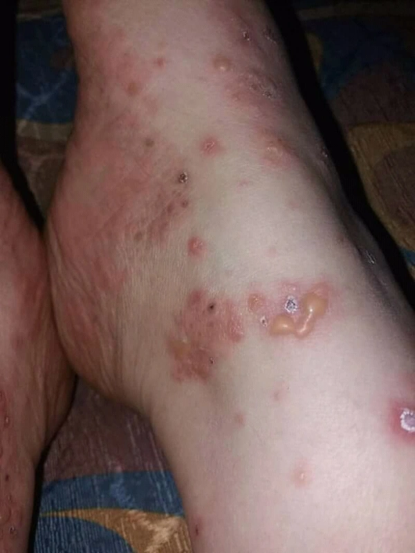
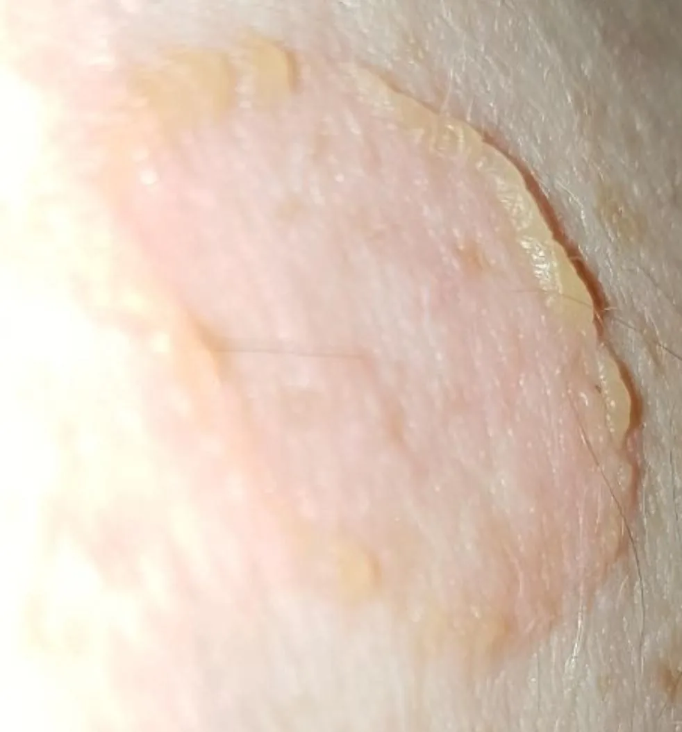
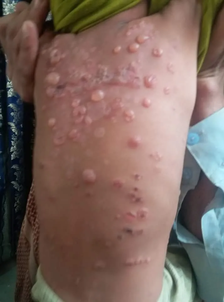
Definition
- Pathogenesis: IgA autoantibodies against LAD1 (BP180 ectodomain).
- Clinical features: Affects children (mostly preschool age, mean ~4.5 years) and adults (mainly sixth decade). Characteristic are annularly arranged tense blisters (“string of beads”).
- Special features: frequent mucosal involvement (mouth in about 50 %, also eyes with risk of scarring), pruritus common with variable intensity, skin usually heals without scarring. An important differential diagnosis is TEN (Toxic epidermal necrolysis).
Aetiopathogenesis
Besides idiopathic forms, linear IgA disease can be triggered by drugs; vancomycin is the classic trigger, beta-lactams and NSAIDs are also described. The drug-induced form usually subsides once the culprit is no longer taken.
Diagnosis
- Direct immunofluorescence of perilesional skin: linear IgA deposits along the basement membrane zone.
- Histology: light microscopy is often not definitive and may resemble dermatitis herpetiformis, which shows granular rather than linear IgA deposits on immunofluorescence.
- Serology: detection of circulating IgA antibodies by indirect immunofluorescence, helpful especially when a biopsy is not possible; plus a thorough drug history.
Differential diagnoses
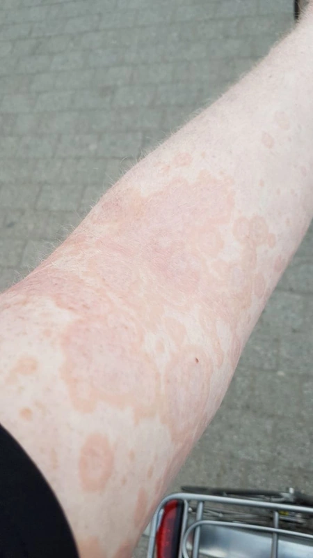
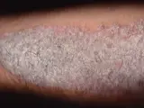 Psoriasis vulgaris (plaque psoriasis)
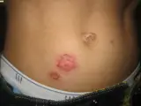 Impetigo (impetigo contagiosa)
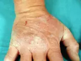 Dermatitis herpetiformis (Duhring disease)
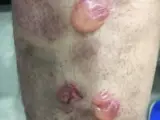 Bullous pemphigoid
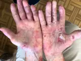 Epidermolysis bullosa acquisita
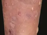 Lichen planus pemphigoides
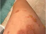 Pemphigoid gestationis
Image credits for the thumbnails
- Psoriasis vulgaris (plaque psoriasis): MediaJet, “An Arm Covered With Plaque Type Psoriasis”, CC BY-SA 3.0, via Wikimedia Commons · CC BY-SA 3.0 · Source
- Impetigo (impetigo contagiosa): Littlekidsdoc, “Bullous impetigo”, CC BY-SA 4.0, via Wikimedia Commons · CC BY-SA 4.0 · Source
- Dermatitis herpetiformis (Duhring disease): BallenaBlanca, “Dermatitis Herpetiforme 2”, CC BY-SA 3.0, via Wikimedia Commons · CC BY-SA 3.0 · Source
- Bullous pemphigoid: Mohammad2018, “Bullous pemphigoid new image”, CC BY-SA 4.0, via Wikimedia Commons · CC BY-SA 4.0 · Source
- Epidermolysis bullosa acquisita: Szymański, Kowalewski, Pietrzyk et al. (2023), “Case Report: Biological treatment of epidermolysis bullosa acquisita: report on four cases and literature review”, CC BY 4.0, via Wikimedia Commons · CC BY 4.0 · Source
- Lichen planus pemphigoides: Gandhi V, Kumar P, Chauhan A (An Bras Dermatol 2022), CC BY 4.0, via PubMed Central (PMC9582882) · CC BY 4.0 · Source · cropped
- Pemphigoid gestationis: Chen RE, Yokoyama CC, Anadkat MJ (JAAD Case Rep 2023), CC BY 4.0, via PubMed Central (PMC10568228) · CC BY 4.0 · Source
Treatment principles
- In drug-induced forms, withdrawing the trigger is crucial; the lesions then usually regress.
- First-line treatment of the idiopathic form is neutrophil-inhibiting systemic therapy, alternatively or additionally systemic glucocorticoids and certain antibiotics, together with topical glucocorticoids.
- Mucosal, and particularly ocular, involvement requires interdisciplinary care; the childhood form often remits spontaneously.
Specific treatment regimens with drugs and doses are covered in the DermaFuchs app.
Keep learning in the app
References (selection)
Cross-references
Mentioned in
Keep learning
Keep learning with flashcards, exam questions and spot diagnoses in the DermaFuchs app – free, in your browser or as an app.
Related pages: Psoriasis vulgaris (plaque psoriasis) · Impetigo (impetigo contagiosa) · Dermatitis herpetiformis (Duhring disease)
Note: This page is intended for medical education and does not replace diagnosis or treatment decisions in individual cases.