Scrofuloderma (Tuberculosis cutis colliquativa)
- Synonyms
- Tuberculosis cutis colliquativa, scrofuloderma
- Specialty
- Dermatology · Bacterial infections
- Images
- Clinical 2
- In the app
- 2 flashcards
- Last updated
- 10/2026 · Dr. Pascal Bafteh
Contents
Images (2)
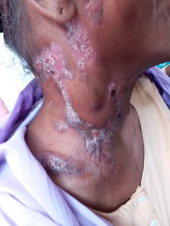
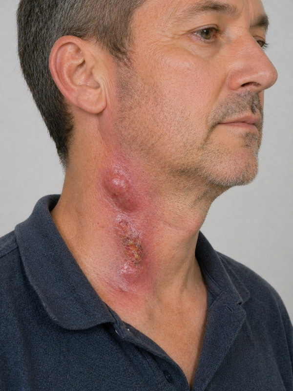 AI illustration
Definition
Scrofuloderma (tuberculosis cutis colliquativa) is a form of cutaneous tuberculosis in which infection spreads to the skin from an underlying tuberculous focus – a lymph node or bone, often with concomitant pulmonary tuberculosis; most commonly around the neck and under the jawline. Firm, painless lesions develop that ulcerate with a granular base.
- Clinical features: presents as subcutaneous fistulating nodules with livid-violaceous raised border and seropurulent secretion in the submandibular, supraclavicular, inguinal, retroauricular regions.
- Pathogenesis: Spread by contiguity from tuberculous lymph nodes.
Diagnosis
- Biopsy with histology (caseating granulomas), Ziehl-Neelsen stain, culture and PCR from tissue.
- Interferon-gamma release assay, chest X-ray and ultrasound or cross-sectional imaging of the underlying focus.
Differential diagnoses
 Cutaneous myiasis
 Furuncle and carbuncle
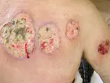 Pyoderma gangrenosum
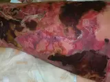 Necrotizing fasciitis
 Hidradenitis suppurativa (acne inversa)
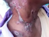 Cutaneous tuberculosis
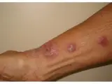 Sporotrichosis
Image credits for the thumbnails
- Cutaneous myiasis: Petruss, “Miasis human”, CC0, via Wikimedia Commons · CC0 · Source
- Furuncle and carbuncle: Medicalpal, “Carbuncle”, CC BY-SA 4.0, via Wikimedia Commons · CC BY-SA 4.0 · Source
- Pyoderma gangrenosum: Monopol, “Pyoderma gangrenosum 01”, CC BY-SA 3.0, via Wikimedia Commons · CC BY-SA 3.0 · Source
- Necrotizing fasciitis: CNX OpenStax, CC BY 4.0, via Wikimedia Commons, cropped · CC BY 4.0 · Source
- Hidradenitis suppurativa (acne inversa): Dr. Thomas Brinkmeier, “Acne inversa, Hurley-Stadium II, ©WIKIDERM”, CC BY 4.0, via Wikimedia Commons · CC BY 4.0 · Source
- Cutaneous tuberculosis: Mohammad2018, “Scrofuloderma”, CC BY-SA 4.0, via Wikimedia Commons · CC BY-SA 4.0 · Source
- Sporotrichosis: Gomes RDSR, do Valle ACF, Freitas DFS et al. (J Fungi (Basel) 2023), CC BY 4.0, via PubMed Central (PMC10455193) · CC BY 4.0 · Source
Treatment principles
- Scrofuloderma is treated like organ tuberculosis, with standard combination antituberculous therapy.
- It is important to look for the underlying focus (e.g. lymph nodes, bone or joints) and to screen contacts; tuberculosis is a notifiable disease.
- Surgical measures such as clearance of necrotic lymph nodes are needed only as an adjunct.
Specific treatment regimens with drugs and doses are covered in the DermaFuchs app.
Keep learning in the app
References (selection)
Cross-references
Keep learning
Keep learning with flashcards, exam questions and spot diagnoses in the DermaFuchs app – free, in your browser or as an app.
Related pages: Cutaneous myiasis · Furuncle and carbuncle · Pyoderma gangrenosum
Note: This page is intended for medical education and does not replace diagnosis or treatment decisions in individual cases.