Calciphylaxis
- Synonyms
- Calciphylaxie, calcific uraemic arteriolopathy
- Specialty
- Dermatology · Vascular skin disorders
- Images
- Clinical 2
- In the app
- 7 flashcards
- Last updated
- 10/2026 · Dr. Pascal Bafteh
Contents
Images (2)
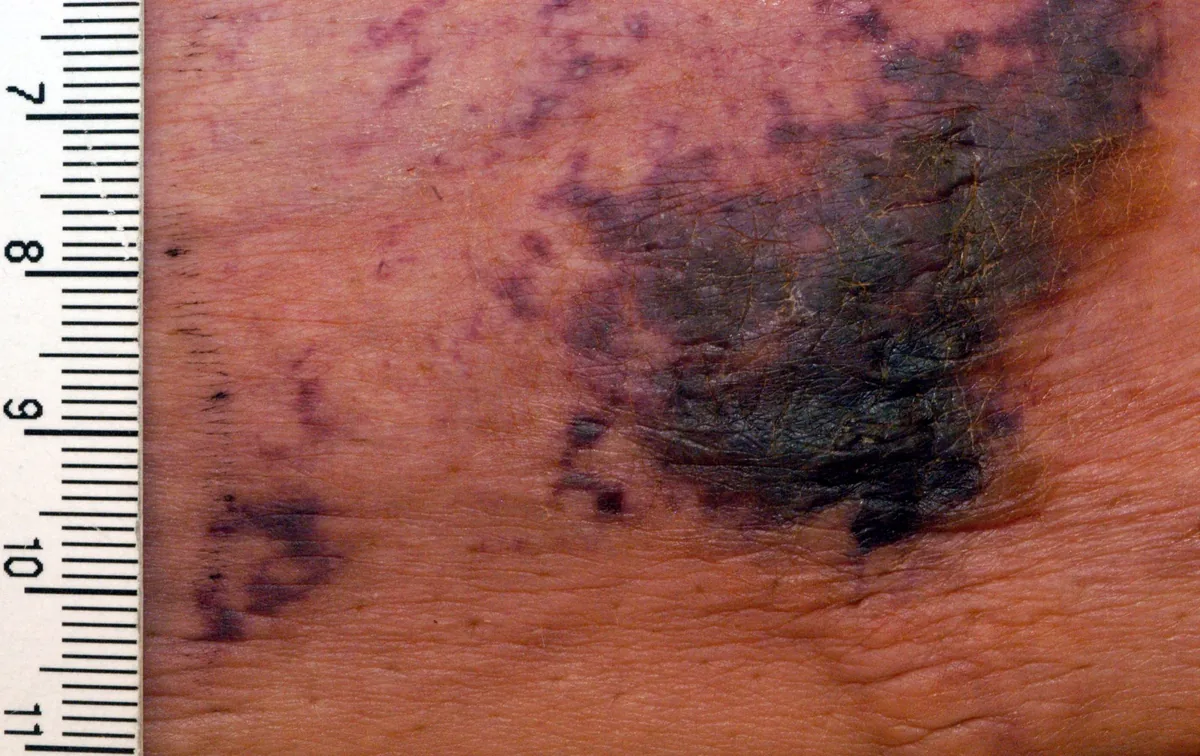
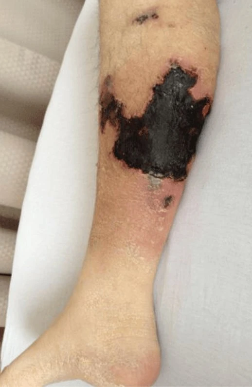
Definition
Definition & Synonyms
- Calciphylaxis (CUA, Calcific Uremic Arteriolopathy) = life-threatening microvascular calcinosis with medial calcification of dermal and subcutaneous arterioles + skin ischemia.
- Synonyms: Synonyms are uremic gangrene + calcific uremic arteriolopathy.
- Incidence in dialysis patients: depending on the survey approx. 0.04–1 % per year; 1-year mortality 45–80 % (sepsis from wound infection!).
Aetiopathogenesis
Pathogenesis
- Imbalance of pro-/anti-calcification factors: Key mechanism is vit-K deficiency + Fetuin-A ↓ + Matrix-Gla protein inactive plus hyperphosphatemia + secondary hyperparathyroidism.
- Microangiopathy: Media calcification → intimal proliferation → microthrombi → ischemic necrosis.
- Risk factors: Common RF are dialysis requirement + obesity + diabetes + female sex + vit-K antagonists.
Clinical features
Clinical Presentation
- Early phase: Livedoid reddish-livid macules / plaques, very painful (even without visible lesion: "pain before skin finding").
- Late phase: Typical are sharply demarcated black ulcers with hard eschar-like necrosis + livid border on adipose locations: inner thigh / abdomen / breasts / gluteal.
- Special form "distal": on acral areas — DD PAD, cholesterol embolism.
- Extremely painful — often opioid-requiring.
Diagnosis
Diagnostics
- Laboratory often unremarkable: According to Braun-Falco (p. 1715), there are no or only minor changes in PTH and Ca×P, no pathognomonic laboratory values. Ca×P > 70, ↑ PTH or ↑ phosphate are at most hints/risk factors, NOT diagnostic criteria — the diagnosis is primarily clinical-histopathological (livedoid plaques + ischemic necroses + media sclerosis with calcification on histology).
- Skin biopsy from border: Diagnostic gold standard is punch / spindle biopsy with Von Kossa stain → media calcification of dermal arterioles + microthrombi + cutaneous necrosis.
- Imaging (optional): plain X-ray with vascular calcifications, high-resolution US, mammography as a supplement when the breast is involved.
Differential diagnoses
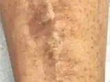
Image credits for the thumbnails
- Pyoderma gangrenosum: Monopol, “Pyoderma gangrenosum 01”, CC BY-SA 3.0, via Wikimedia Commons · CC BY-SA 3.0 · Source
- Necrotizing fasciitis: CNX OpenStax, CC BY 4.0, via Wikimedia Commons, cropped · CC BY 4.0 · Source
- Diabetic dermopathy (shin spots): Abate, Aroucha, Nóbrega et al. (2025), “Cutaneous manifestations of diabetes mellitus: a narrative review”, CC BY 4.0, via Wikimedia Commons · CC BY 4.0 · Source
Treatment principles
- Management is interdisciplinary (nephrology, dermatology, wound care, pain management).
- Modifiable triggers are eliminated immediately, particularly vitamin K antagonists, calcium-containing phosphate binders and vitamin D preparations; calcium-phosphate metabolism is optimised, if necessary with calcimimetics or parathyroidectomy.
- Wound care with cautious debridement, consistent analgesia and infection control are central, as sepsis is the most common cause of death.
Specific treatment regimens with drugs and doses are covered in the DermaFuchs app.
Keep learning in the app
References (selection)
Cross-references
Mentioned in
Keep learning
Keep learning with flashcards, exam questions and spot diagnoses in the DermaFuchs app – free, in your browser or as an app.
Related pages: Arterial (ischaemic) ulcer · Pyoderma gangrenosum · Necrotizing fasciitis
Note: This page is intended for medical education and does not replace diagnosis or treatment decisions in individual cases.