Reactive arthritis (Reiter syndrome)
- Synonyms
- reaktive Arthritis, reactive arthritis, Reiter's syndrome
- Specialty
- Dermatology · Inflammatory skin diseases
- Images
- Clinical 1
- In the app
- 2 flashcards
- Last updated
- 10/2026 · Dr. Pascal Bafteh
Contents
Images (1)
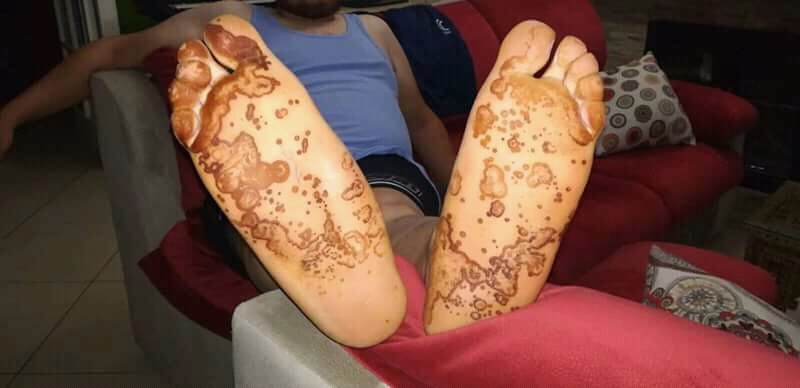
Definition
Definition & Classification
- Classification: seronegative spondyloarthritis, often with psoriasiform skin and nail findings.
Aetiopathogenesis
Etiopathogenesis
- HLA Association: HLA-B27 in up to ~80% of patients.
- Mechanism: infectious-allergic reaction with cross-reaction to HLA-B27.
- Common Trigger Pathogens:
- Urethritic: Chlamydia trachomatis, Mycoplasma genitalium, Ureaplasma
- Enteritic: Yersinia enterocolitica, Salmonella, Shigella, Campylobacter jejuni
- Viral: HIV (Reiter's described as an HIV manifestation)
Classic Triad
- Classic Triad: Arthritis + conjunctivitis + urethritis (Can't see, can't pee, can't climb a tree).
Arthritis
- Clinical Presentation: asymmetric, primarily lower extremities (knees, ankles, feet), often oligoarticular.
- Spondyloarthritis-Typical Associated Manifestations:
- Enthesitis (tendon/ligament insertion inflammation, classically Achilles tendon / plantar fascia)
- Dactylitis ("sausage finger" / "sausage toe" — diffuse swelling of an entire finger/toe)
- Sacroiliitis with back pain, morning stiffness
Clinical features
Skin Manifestations
- Balanitis circinata: annular-erosive plaques on the glans penis — highly characteristic.
- Keratoderma blennorrhagicum: psoriasiform hyperkeratosis on palms and soles.
- Psoriasiform plaques on trunk and extremities possible (psoriasis-like).
- Aphthae / oral involvement: painless mucosal ulcers on the palate / buccal mucosa.
- Onychodystrophy / Onycholysis similar to psoriatic nail.
Diagnosis
Diagnostics
- History of preceding infection (1–4 weeks before onset).
- Laboratory: ESR / CRP ↑, ANA / RF / CCP negative (seronegative!), HLA-B27 (positive in up to ~80%).
- Urethral / Stool Pathogen Detection: Urethral swab + PCR (Chlamydia trachomatis), stool PCR / culture.
- Imaging: Joint ultrasound (effusion), MRI in case of suspected sacroiliitis, X-ray classically shows erosive changes only late.
- Ophthalmological consultation in case of suspected uveitis.
Differential diagnoses
 Psoriatic arthritis
 Secondary syphilis (lues II)
 Hand, foot and mouth disease
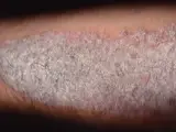 Psoriasis vulgaris (plaque psoriasis)
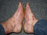 Palmoplantar psoriasis
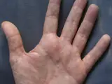 Dyshidrotic eczema (pompholyx)
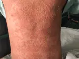 Generalized pustular psoriasis (von Zumbusch)
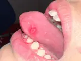 Behçet's disease
Image credits for the thumbnails
- Psoriatic arthritis: James Heilman, MD, “Psoriatic arthritis2010”, CC BY-SA 3.0, via Wikimedia Commons · CC BY-SA 3.0 · Source
- Secondary syphilis (lues II): Ehlers S, Sergent S, Ashurst J (Clin Pract Cases Emerg Med 2020), CC BY 4.0, via PubMed Central (PMC7676796) · CC BY 4.0 · Source
- Hand, foot and mouth disease: Kessalia19, CC BY-SA 4.0, via Wikimedia Commons · CC BY-SA 4.0 · Source
- Psoriasis vulgaris (plaque psoriasis): MediaJet, “An Arm Covered With Plaque Type Psoriasis”, CC BY-SA 3.0, via Wikimedia Commons · CC BY-SA 3.0 · Source
- Palmoplantar psoriasis: MCvanVeen, “Psoriasis July 2016”, CC BY-SA 4.0, via Wikimedia Commons · CC BY-SA 4.0 · Source
- Dyshidrotic eczema (pompholyx): Inwe (Wikimedia Commons), “Dyshidrosis”, CC BY-SA 3.0, via Wikimedia Commons · CC BY-SA 3.0 · Source
- Generalized pustular psoriasis (von Zumbusch): Bourousis E, Kotzamani MT, Maritsi DN., Cureus 2025, Figure 1, CC BY 4.0, via PMC · CC BY 4.0 · Source
- Behçet's disease: El Omari S, Barhoud O, Elarabi S et al. (Cureus 2026), CC BY 4.0, via PubMed Central (PMC13561507) · CC BY 4.0 · Source · cropped
Treatment principles
- The triggering infection is treated with antibiotics, including treatment of partners in urogenital chlamydial infection.
- Arthritis is first treated with non-steroidal anti-inflammatory drugs and, where appropriate, intra-articular glucocorticoids; chronic disease is managed with disease-modifying drugs and biologics.
- Skin and mucosal lesions are treated topically, and uveitis by an ophthalmologist.
Specific treatment regimens with drugs and doses are covered in the DermaFuchs app.
Keep learning in the app
Cross-references
Mentioned in
Keep learning: test yourself
One question from the DermaFuchs app on Reactive arthritis (Reiter syndrome) – tap an answer.
Question 1 of 1
What is the most common initial ocular finding in Reiter's syndrome (reactive arthritis) that can later progress to anterior uveitis?
Answer
Correct: bilateral conjunctivitis. Bilateral conjunctivitis is a frequent and early ocular manifestation of reactive arthritis, which may sometimes progress to more severe inflammation like anterior uveitis.
More questions like these – with explanations and spaced repetition – in the app.
Note: This page is intended for medical education and does not replace diagnosis or treatment decisions in individual cases.