Eosinophilic dermatoses
Exam relevance: in 1 of more than 50 board exam reports · rank 93
- Synonyms
- eosinophilic skin diseases
- Specialty
- Dermatology · Inflammatory skin diseases
- Images
- Clinical 5 · Histology 1
- In the app
- 2 flashcards
- Last updated
- 10/2026 · Dr. Pascal Bafteh
Contents
Images (6)
 Histology
Definition
Overview: Eosinophil-driven Dermatoses
| Disease | Pathogenesis (Brief) | Characteristic |
|---|---|---|
| Wells Syndrome (Eosinophilic Cellulitis) | Reaction to various triggers; MBP release + Leukotrienes | Flame Figures in histo |
| Papuloerythroderma Ofuji (PEO) | unclear, rarely paraneoplastic / older age | Deck-Chair Sign, trunk-flexural sparing |
| Eosinophilic Pustulosis Ofuji (EPF) | follicular-eosinophilic | sterile follicular pustules, face / trunk |
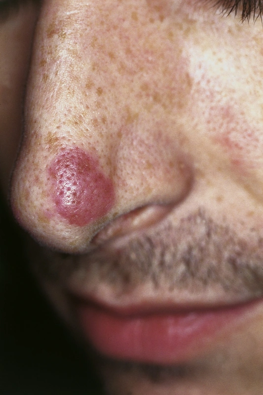
| Granuloma faciale | unclear, Th2 + LCV variant | white men 40–60 yrs, almost only face |
| Eosinophilic Dermatitis Nir-Westfried | unclear, skin-limited HES variant | dissem. itchy papules, eosinophilia > 10 % |
| Eosinophilic Fasciitis (Shulman) | immune-mediated, partly after exertion | Mattress Phenomenon, profound sclerosis |
| Hypereosinophilic Syndrome (HES) | clonal (PDGFRA) or reactive | Eosinophils ≥ 1500/µl on repeated counts + organ involvement |
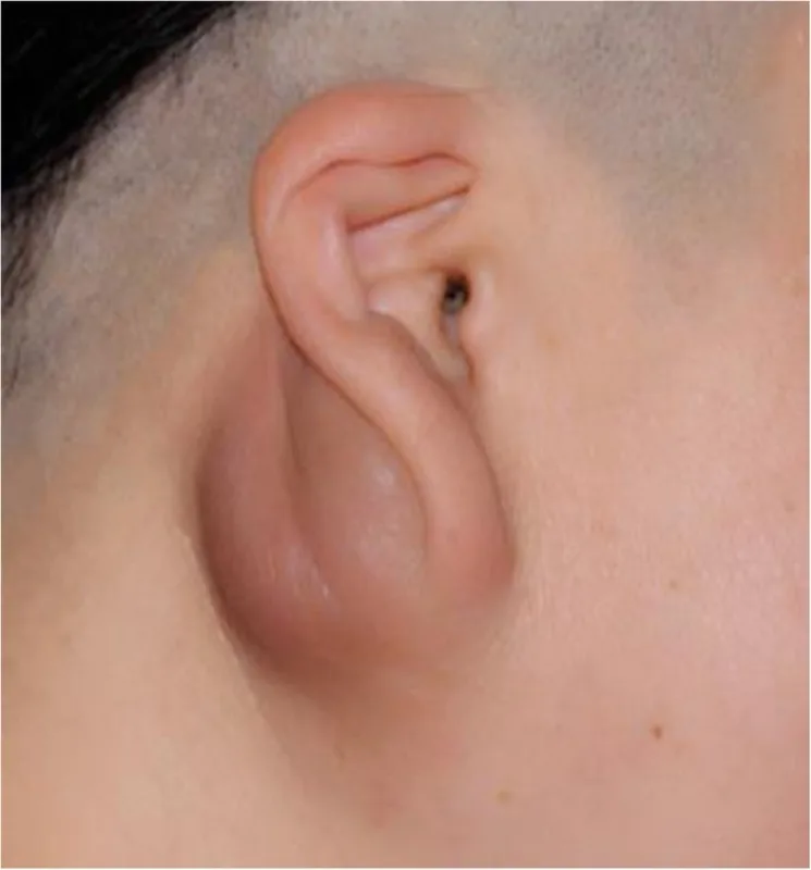
| Kimura Disease / ALHE | reactive-vascular | Lymph node swelling, IgE↑, ALHE = superficial |
Classification
Papuloerythroderma Ofuji (PEO)
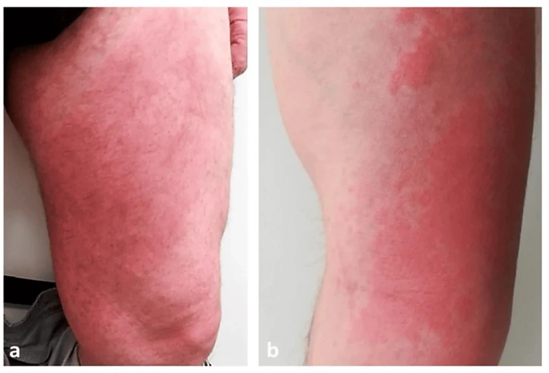
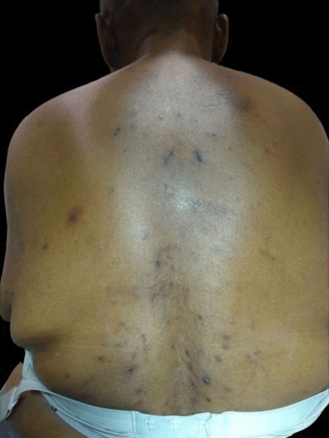
- Clinical Features: Characteristic is a maculopapular rash with significant pruritus and confluence leading to erythroderma.
- Localization Key Finding: Characteristic is the sparing of large flexures as Deck-Chair Sign, face / palms / soles free.
- Laboratory: Blood eosinophilia, elevated IgE, lymphopenia common.
- Association: malignancies, atopy, infections or drugs are only rarely associated; a cutaneous T-cell lymphoma may underlie it histologically — hence work-up including biopsy.
Eosinophilic Pustulosis Ofuji (EPF)
- NOT to be confused with Papuloerythroderma Ofuji!
- Clinical Features: Characteristic are sterile follicular pustules with an annular arrangement on the face / trunk, mostly adult Asian men (classic type).
- Histology: eosinophilic spongiosis + folliculitis with eosinophilic microabscesses.
Granuloma faciale
- Clinical Features: Typical are white men 40–60 yrs + face with brownish-livid plaques and peau d'orange appearance.
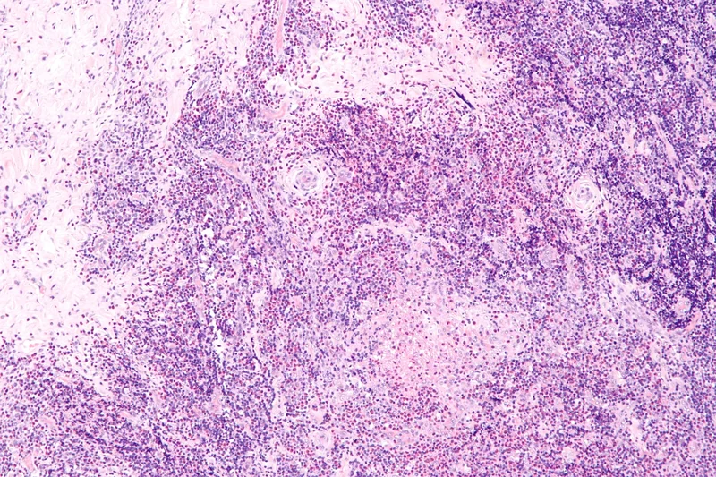
- Histology: Characteristic is a polymorphic eosinophilic infiltrate with leukocytoclastic vasculitis and subepidermal Grenz zone.
Eosinophilic Fasciitis (Shulman Syndrome)
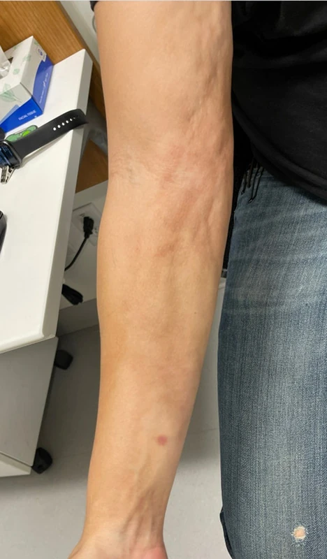
- Clinical Features: Characteristic: deep scleroderma-like induration, partly after exertion; initially oedema, later orange-peel skin (mattress phenomenon) and groove sign.
- Sparing Sign: Hands / feet usually free (vs. SSc!), Raynaud's phenomenon typically negative.
- Laboratory: Blood eosinophilia, ↑ESR / CRP, hypergammaglobulinemia, ANA / Scl-70 negative.
- Diagnostics: MRI of the fascia (contrast enhancement), spindle biopsy down to fascia / muscle with evidence of eosinophilic fasciitis + fibrosis.
Hypereosinophilic Syndrome (HES) — brief
- Definition: HES is defined as Eosinophilia ≥ 1 500/µl on repeated counts with eosinophil-related organ involvement (heart, lung, skin, GI tract, CNS); classically persisting ≥ 6 months, today ≥ 2 counts at least 2 weeks apart suffice.
- Skin manifestations: Pruritus + urticarial plaques + erythematous papules + rarely mucosal ulcers.
Keep learning in the app
References (selection)
Cross-references
Keep learning
Keep learning with flashcards, exam questions and spot diagnoses in the DermaFuchs app – free, in your browser or as an app.
Related pages: Eosinophilic fasciitis (Shulman) · Erythema annulare centrifugum (EAC) · Erythema nodosum
Note: This page is intended for medical education and does not replace diagnosis or treatment decisions in individual cases.