Pyogenic granuloma
Exam relevance: in 3 of more than 50 board exam reports · rank 45
- Synonyms
- pyogenes Granulom, lobular capillary haemangioma, bleeding red bump
- Specialty
- Dermatology · Skin tumours
- Images
- Clinical 6 · Histology 1
- In the app
- 4 flashcards
- Last updated
- 10/2026 · Dr. Pascal Bafteh
Contents
Images (7)
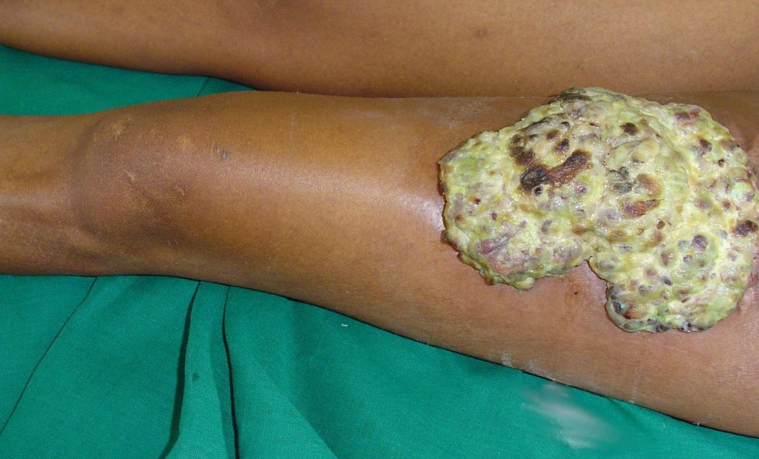
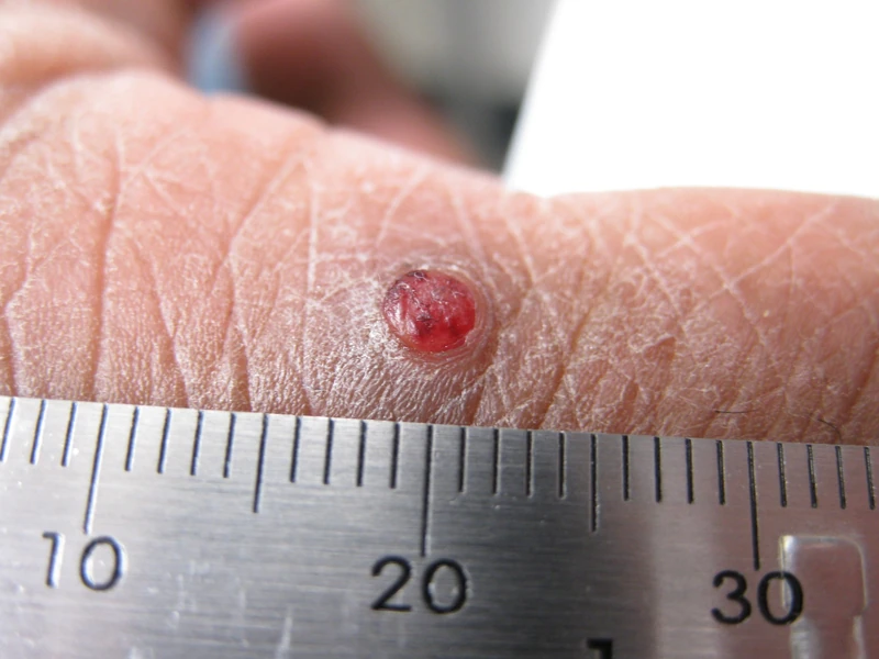
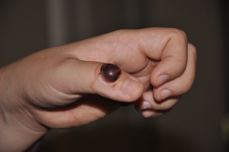
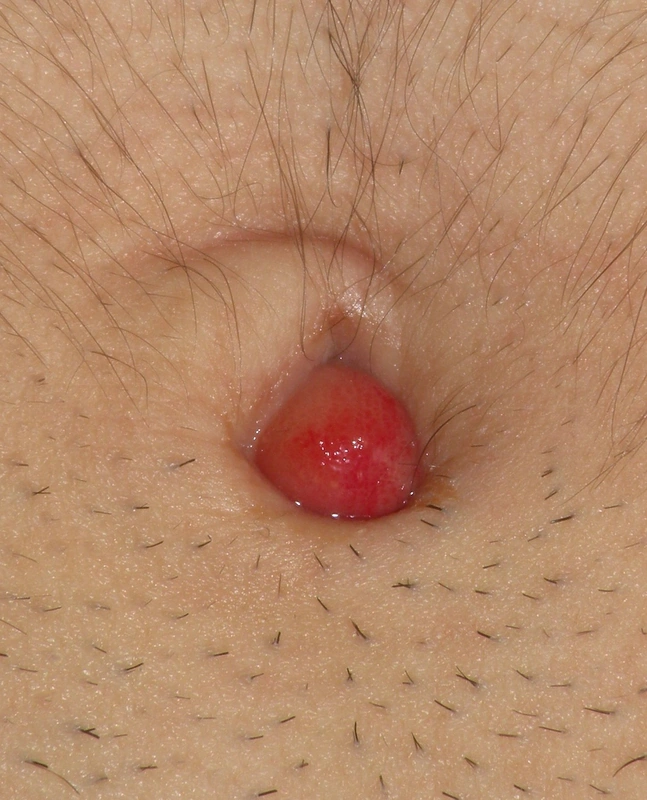
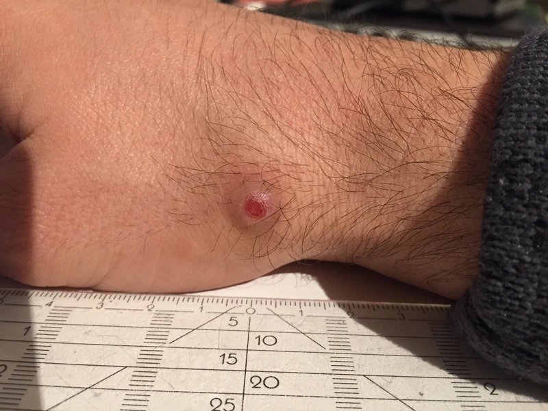
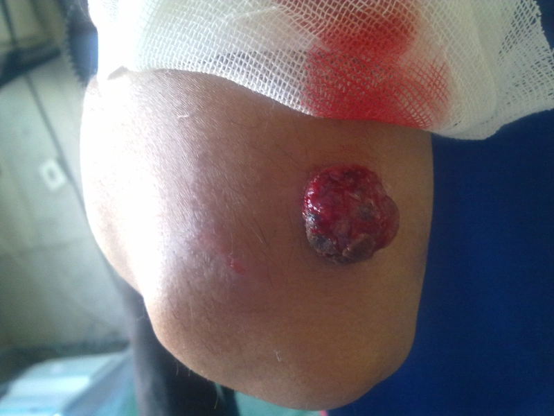
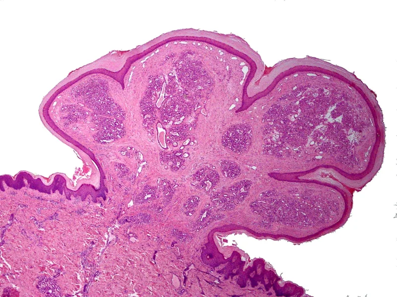 Histology
Definition
- Classification: Despite its name neither purulent nor granulomatous, but a vascular tumour in the ISSVA classification — histologically a lobular capillary hemangioma with lobular capillary sprouts in an oedematous stroma.
- Key clinical picture: A rapidly growing, soft, bright red, easily bleeding nodule appearing within weeks.
- Morphology: usually under 1–2 cm, pedunculated or sessile, often eroded and crusted.
- At the base: frequently an epidermal collarette.
- Characteristic: profuse bleeding tendency on minimal contact — patients often present because of repeated bleeding that is hard to stop.
- The critical differential diagnosis: Amelanotic melanoma can look clinically identical — rapidly growing, red, bleeding, on an acral site.
- Further differentials: angiosarcoma (scalp, older patients) · Kaposi sarcoma · bacillary angiomatosis under immunosuppression · ulcerated basal or squamous cell carcinoma · in children Spitz naevus.
- Consequence: no pyogenic granuloma without histological work-up — never simply cauterise and discard.
- Dermoscopy: homogeneous reddish to red-white areas with whitish separating lines ("white rail lines") and a peripheral collarette; melanocytic criteria are absent. This supports the clinical suspicion but does not replace histology, because amelanotic melanoma can mimic the same pattern.
Differential diagnoses
 Melanoma
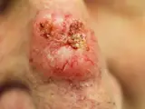 Basal cell carcinoma
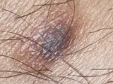 Dermatofibroma
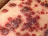 Kaposi's sarcoma
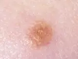 Spitz naevus (spindle and epithelioid cell naevus)
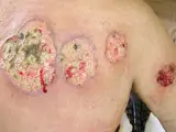 Pyoderma gangrenosum
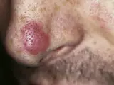 Granuloma faciale
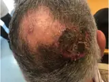 Cutaneous angiosarcoma
Image credits for the thumbnails
- Melanoma: National Cancer Institute (unknown photographer), “Melanoma (4)”, Public Domain, via Wikimedia Commons · Public domain · Source
- Basal cell carcinoma: Bin im Garten, “Basaliom am Nasenrücken 2”, CC BY-SA 3.0, via Wikimedia Commons · CC BY-SA 3.0 · Source
- Dermatofibroma: Jmarchn, “Pigmented and tenderness dermatofibroma”, CC BY-SA 3.0, via Wikimedia Commons · CC BY-SA 3.0 · Source
- Kaposi's sarcoma: unknown author, “Kaposi's Sarcoma”, Public Domain, via Wikimedia Commons · Public Domain · Source
- Spitz naevus (spindle and epithelioid cell naevus): The Armed Forces Institute of Pathology (AFIP), Public domain, via Wikimedia Commons · Public domain · Source
- Pyoderma gangrenosum: Monopol, “Pyoderma gangrenosum 01”, CC BY-SA 3.0, via Wikimedia Commons · CC BY-SA 3.0 · Source
- Granuloma faciale: unknown (Commons user), “Facial eosinophilic granuloma 01”, CC BY 2.0, via Wikimedia Commons · CC BY 2.0 · Source
- Cutaneous angiosarcoma: Messias H, Martins M, Zagalo C et al. (Cancer Rep (Hoboken) 2022), CC BY 4.0, via PubMed Central (PMC9575495) · CC BY 4.0 · Source
Treatment principles
- Treatment usually consists of removal, for example by curettage or shave excision with electrocoagulation of the base, or by excision; the tissue is always examined histologically, particularly to exclude amelanotic melanoma.
- Laser treatment or cryotherapy are alternatives.
- Recurrence is common after incomplete removal; lesions arising in pregnancy may regress spontaneously after delivery, and drug-induced lesions after withdrawal of the causative drug.
Specific treatment regimens with drugs and doses are covered in the DermaFuchs app.
Keep learning in the app
References (selection)
Cross-references
Mentioned in
Keep learning
Keep learning with flashcards, exam questions and spot diagnoses in the DermaFuchs app – free, in your browser or as an app.
Related pages: Melanoma · Basal cell carcinoma · Dermatofibroma
Note: This page is intended for medical education and does not replace diagnosis or treatment decisions in individual cases.