Leg ulcer (ulcus cruris)
Exam relevance: in 11 of more than 50 board exam reports · rank 7
- Synonyms
- offenes Bein, Unterschenkelgeschwür, Ulcus cruris venosum, Ulcus cruris arteriosum, leg ulcer, venous leg ulcer, venous ulcer, chronic wound
- Specialty
- Dermatology · Vascular skin disorders
- Images
- Clinical 10
- In the app
- 4 flashcards
- Last updated
- 10/2026 · Dr. Pascal Bafteh
Contents
Images (10)
Definition
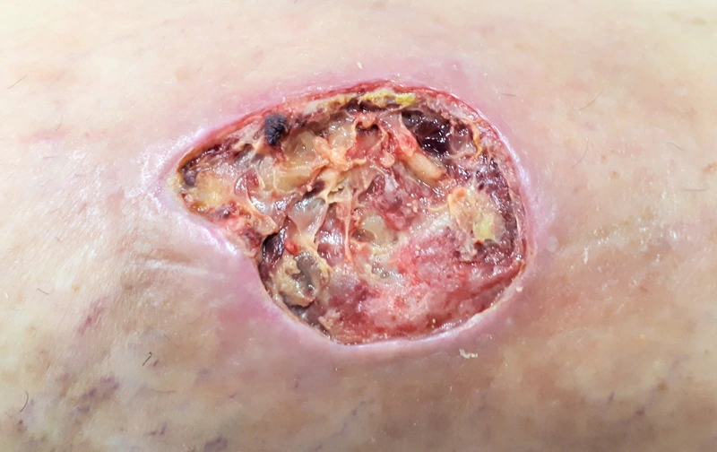
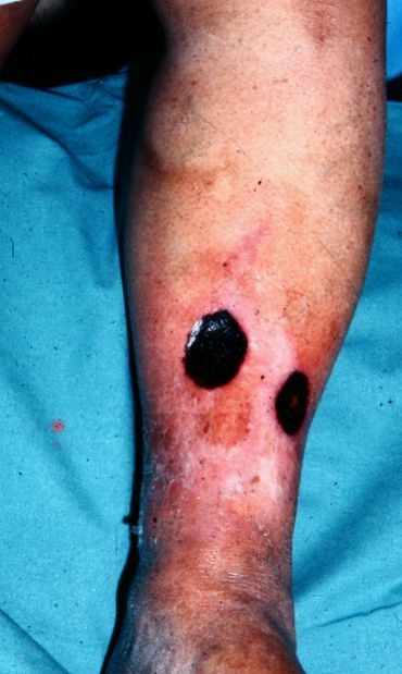
A leg ulcer is a full-thickness skin defect on the lower leg or foot, whatever the cause. An ulcer is considered chronic if it persists for longer than 4 weeks. Venous, arterial or neuropathic disease is the most common underlying cause; a minor injury or pressure is often the trigger.
Occurrence & epidemiology
Chronic leg ulcers affect about 1% of middle-aged and elderly people; in Germany more than 1 million people are affected. The distribution of causes depends strongly on the population studied:
- venous (chronic venous insufficiency): 45–80%
- arterial (peripheral arterial disease): 5–20%
- diabetic (arterial insufficiency and neuropathy): 15–25%
- In a German dermatological wound centre (354 patients), 57% were venous, 15% mixed arterial-venous, 13% vasculitic and 3.7% purely arterial; 16 causes were identified overall.
- In an analysis of 1000 patients from the same centre, about 70% of causes were vascular and 30% rare (eg, pyoderma gangrenosum, necrobiosis lipoidica, which are relatively more common in younger patients).
Aetiopathogenesis
- Venous: valve incompetence, venous obstruction or impaired calf muscle pump cause venous hypertension with pericapillary fibrin deposits. Risk factors include varicose veins, previous deep vein thrombosis (post-thrombotic syndrome), leg swelling, prolonged standing or sitting, multiple pregnancies, obesity and immobility.
- Arterial: mostly atherosclerosis with reduced blood supply; risk factors are smoking, diabetes, hypercholesterolaemia, hypertension, renal failure and other vascular disease.
- Diabetic: a combination of arterial insufficiency and sensory neuropathy, so that pressure and injuries go unnoticed.
- Less common causes: vasculitis, pyoderma gangrenosum, systemic sclerosis, skin cancer, infection (eg, ecthyma) and pressure.
Clinical features
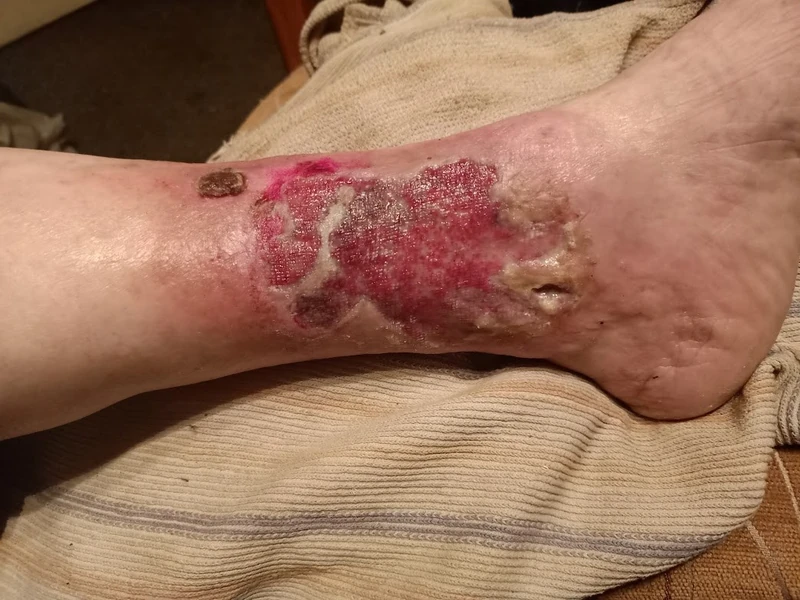
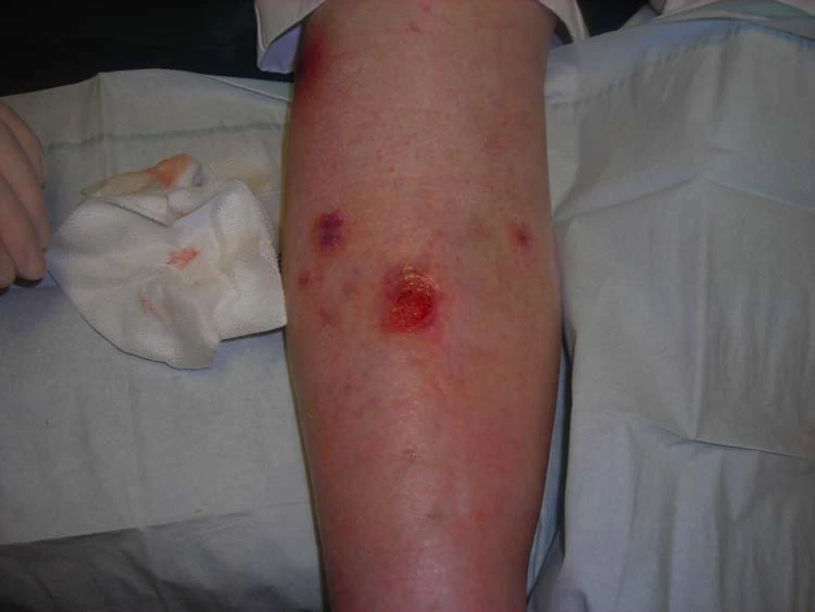
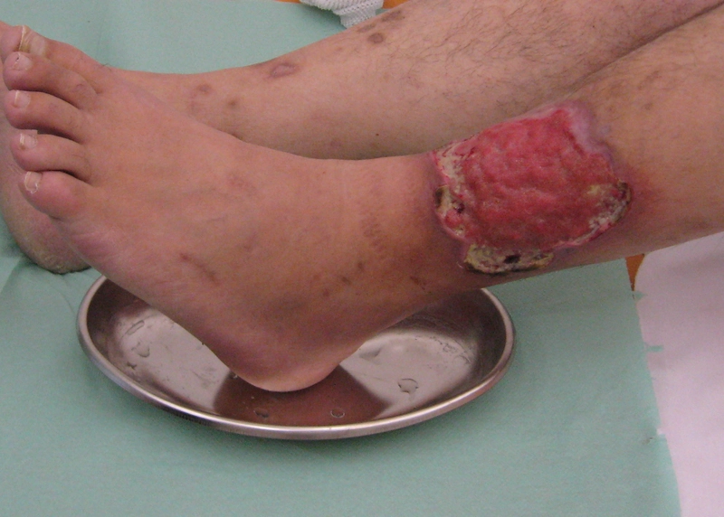
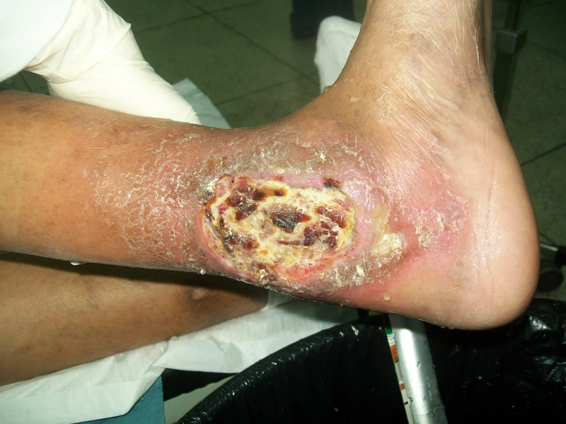
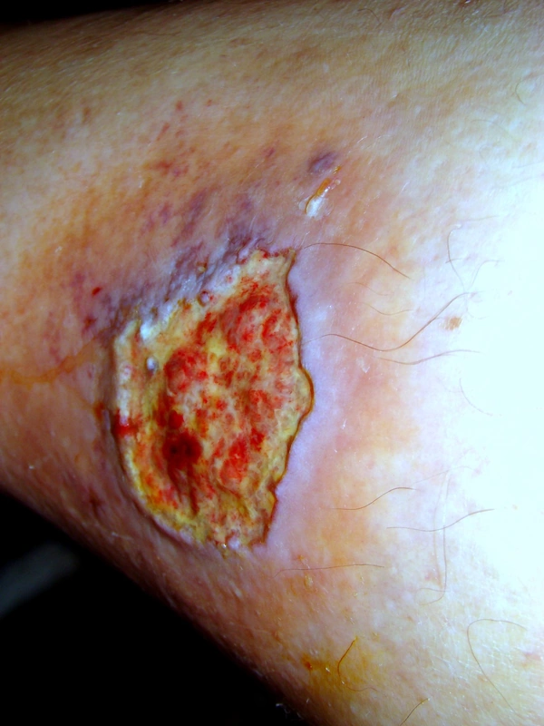
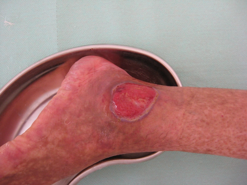
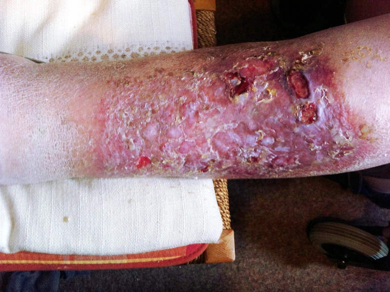
- Venous ulcer: below the knee, most often at the medial ankle; shallow and moist, relatively painless unless infected; surrounding oedema, red-brown staining, venous eczema and lipodermatosclerosis, often atrophie blanche as a scar; heavy, aching legs that feel better when elevated.
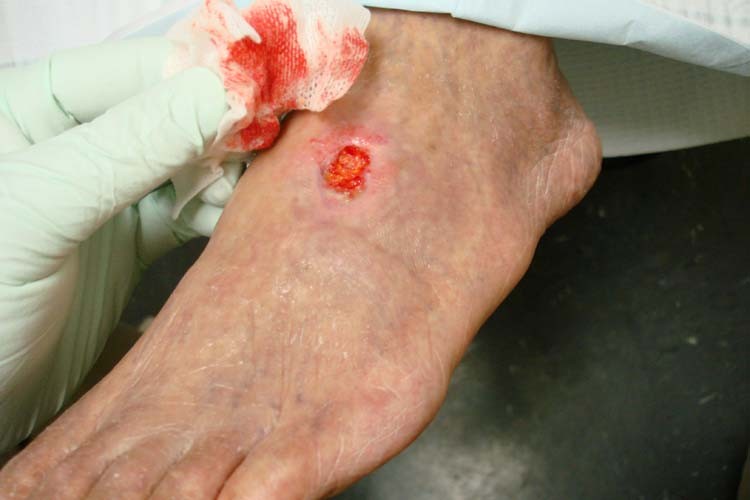
- Arterial ulcer: on the feet, heels and toes, punched-out; painful especially at night and with the legs elevated, relieved by lowering the legs; cold, white or bluish, shiny skin, intermittent claudication; deep ulcers may expose tendons or bone.
- Diabetic ulcer: over pressure points (heel, tips of the toes, between the toes), often with callus.
- Infected ulcer: yellowish crust or pus, unpleasant smell, tender redness, warmth and swelling of the surrounding skin.
Diagnosis
- History (vascular risk factors, thrombosis, injury, type of pain), inspection and pulse examination.
- Ankle-brachial index (ABI) using a Doppler probe: 0.90 or less is diagnostic of peripheral arterial disease, 0.91–0.99 is borderline, 1.00–1.40 normal; values below 0.5 indicate severe arterial disease. Values above 1.40 suggest non-compressible, calcified arteries (Mönckeberg medial sclerosis); if suspicion persists, toe pressure and the toe-brachial index are then measured.
- Duplex ultrasound of the leg veins when chronic venous insufficiency is suspected and to exclude deep vein thrombosis.
- Biopsy from the ulcer edge if suspicious, to confirm or exclude skin cancer.
Treatment principles
- Investigating and treating the cause is essential: in venous ulcers, compression therapy (after excluding significant peripheral arterial disease) and treatment of varicose veins are central.
- Local treatment is phase-appropriate, with debridement, moist wound care and infection control; large, non-healing ulcers can be covered by skin grafting.
- Treatment-resistant or atypical ulcers are biopsied so as not to miss malignancy, vasculitis or pyoderma gangrenosum.
Specific treatment regimens with drugs and doses are covered in the DermaFuchs app.
Keep learning in the app
References (selection)
- DermNet: Leg ulcers
- MSD Manual Professional: Peripheral arterial disease
- MSD Manual Professional: Chronic venous insufficiency and postphlebitic syndrome
- [Causes of leg ulcers. Analysis of the data from a dermatologic wound care center]
- [First manifestation of leg ulcers : Analysis of data from 1000 patients]
Cross-references
More vascular skin disorders
- Urticarial vasculitis (normocomplementaemic and hypocomplementaemic)
- Antiphospholipid syndrome – skin manifestations (livedo racemosa, Sneddon syndrome)
- Cholesterol embolism (atheroembolism)
- Diabetic foot ulcer (malum perforans)
- Erythromelalgia
- Granulomatosis with polyangiitis (GPA) – skin manifestations
- IgA vasculitis (Henoch-Schönlein purpura)
- Calciphylaxis
Keep learning
Keep learning with flashcards, exam questions and spot diagnoses in the DermaFuchs app – free, in your browser or as an app.
Related pages: Urticarial vasculitis (normocomplementaemic and hypocomplementaemic) · Antiphospholipid syndrome – skin manifestations (livedo racemosa, Sneddon syndrome) · Cholesterol embolism (atheroembolism)
Note: This page is intended for medical education and does not replace diagnosis or treatment decisions in individual cases.