Granuloma faciale
- Specialty
- Dermatology · Inflammatory skin diseases
- Images
- Clinical 1
- In the app
- 2 flashcards
- Last updated
- 10/2026 · Dr. Pascal Bafteh
Contents
Images (1)
Definition
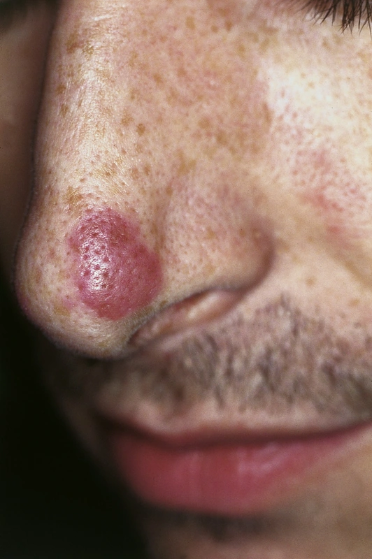
Granuloma faciale is a rare, benign, chronic skin disease with single or multiple, usually reddish-brown papules, plaques or nodules, mostly on the face (rarely extrafacial); middle-aged men are most often affected. Histologically it is a localised leucocytoclastic vasculitis with a mixed dermal infiltrate containing eosinophils and usually a grenz zone beneath the epidermis – the term "granuloma" is misleading.
- Clinical Features: Typical are white men 40–60 yrs + face with brownish-livid plaques and peau d'orange appearance.
- Histology: Characteristic is a polymorphic eosinophilic infiltrate with leukocytoclastic vasculitis and subepidermal Grenz zone.
Diagnosis
- Clinical: soft, raised, well-defined lesions with prominent follicular openings; usually symptomless, occasionally tender, itchy or stinging.
- Biopsy for confirmation; distinction from, among others, erythema elevatum diutinum, sarcoidosis, cutaneous lupus erythematosus, polymorphic light eruption, lymphocytoma cutis, mycosis fungoides and fixed drug eruption.
- A similar change can occur on the mucosa of the upper airways (eosinophilic angiocentric fibrosis).
Differential diagnoses
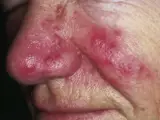 Rosacea
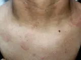 Lupus erythematosus
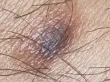 Dermatofibroma
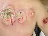 Pyoderma gangrenosum
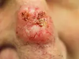 Basal cell carcinoma
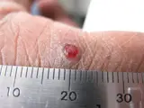 Pyogenic granuloma
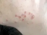 Cutaneous sarcoidosis
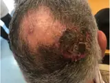 Cutaneous angiosarcoma
Image credits for the thumbnails
- Rosacea: Michael Sand, Daniel Sand, Christina Thrandorf, Volker Paech, Peter Altmeyer, Falk G Bechara, “Rosacea”, CC BY 2.5, via Wikimedia Commons, cropped · CC BY 2.5 · Source
- Lupus erythematosus: Meriam Hajji, CC BY 4.0, via Wikimedia Commons · CC BY 4.0 · Source
- Dermatofibroma: Jmarchn, “Pigmented and tenderness dermatofibroma”, CC BY-SA 3.0, via Wikimedia Commons · CC BY-SA 3.0 · Source
- Pyoderma gangrenosum: Monopol, “Pyoderma gangrenosum 01”, CC BY-SA 3.0, via Wikimedia Commons · CC BY-SA 3.0 · Source
- Basal cell carcinoma: Bin im Garten, “Basaliom am Nasenrücken 2”, CC BY-SA 3.0, via Wikimedia Commons · CC BY-SA 3.0 · Source
- Pyogenic granuloma: kilbad, “Pyogenic granuloma on a finger-1”, CC BY 3.0, via Wikimedia Commons · CC BY 3.0 · Source
- Cutaneous sarcoidosis: Nowack et al., “Cutaneous findings in systemic sarcoidosis”, CC BY-SA 2.0, via Wikimedia Commons · CC BY-SA 2.0 · Source
- Cutaneous angiosarcoma: Messias H, Martins M, Zagalo C et al. (Cancer Rep (Hoboken) 2022), CC BY 4.0, via PubMed Central (PMC9575495) · CC BY 4.0 · Source
Treatment principles
- Granuloma faciale runs a chronic course and is often resistant to treatment; spontaneous regression is rare.
- Topical or intralesional glucocorticoids and topical calcineurin inhibitors are used, as well as physical methods such as cryotherapy or vascular and ablative lasers.
- Excision is possible, but recurrences are common even after surgical removal.
Specific treatment regimens with drugs and doses are covered in the DermaFuchs app.
Keep learning in the app
References (selection)
Cross-references
Mentioned in
Keep learning
Keep learning with flashcards, exam questions and spot diagnoses in the DermaFuchs app – free, in your browser or as an app.
Related pages: Rosacea · Lupus erythematosus · Dermatofibroma
Note: This page is intended for medical education and does not replace diagnosis or treatment decisions in individual cases.