Mucormycosis
- Synonyms
- Zygomykose, Mucormykose, kutane Mukormykose, rhinozerebrale Mukormykose, Zygomycosis, Black fungus
- Specialty
- Dermatology · Fungal infections
- Images
- Clinical 1
- In the app
- 2 flashcards
- Last updated
- 10/2026 · Dr. Pascal Bafteh
Contents
Images (1)
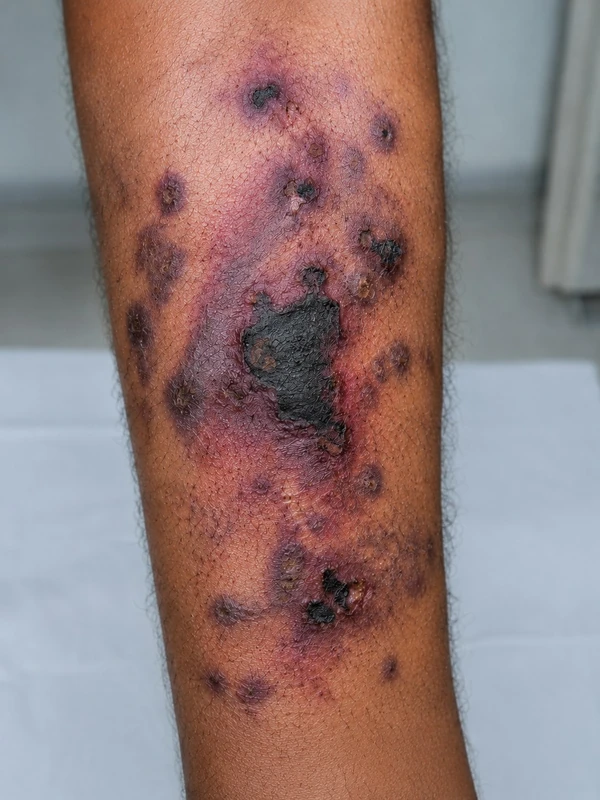 AI illustration
Definition
Mucormycosis is an opportunistic, often rapidly progressive infection caused by moulds of the order Mucorales (mainly Rhizopus, Mucor, Lichtheimia), which are found in soil and decaying plant matter. The organisms invade vessel walls; thrombosis leads to progressive tissue necrosis.
Classification
Rhinocerebral or rhino-orbital-cerebral (34–39%), pulmonary (20–24%), cutaneous (19–22%), gastrointestinal and disseminated.
Occurrence & epidemiology
The leading risk factor worldwide is diabetes mellitus (especially when poorly controlled), followed by haematological malignancies and stem cell transplantation; others include solid organ transplantation, neutropenia and iron overload. Up to 20% of patients are immunocompetent, often after skin injury or burns. The annual incidence ranges from about 1 per million in the US and Europe to 140 per million in India.
Clinical features
- Cutaneous form: primary at the site of entry (wound, burn, tattoo, intravenous line site) or secondary to dissemination. A painful, indurated, erythematous to violaceous plaque that rapidly turns into a black necrotic eschar; insidious or fulminant course up to gangrene.
- Rhino-orbital-cerebral form: necrosis of the nasal mucosa or palate, fever, purulent nasal discharge, swelling around the eye, proptosis, loss of vision; spread to the brain is possible.
Diagnosis
- Confirmation by a deep skin biopsy with tissue for histology and culture.
- Histology: broad, ribbon-like, non-septate hyphae with right-angle branching that invade vessel walls.
- Culture for species identification, negative in up to 50%; PCR for fungal DNA.
Differential diagnoses
Keep learning in the app
References (selection)
Cross-references
Note: This page is intended for medical education and does not replace diagnosis or treatment decisions in individual cases.