Pemphigus vegetans
- Specialty
- Dermatology · Blistering diseases
- Images
- Clinical 1
- In the app
- 4 flashcards
- Last updated
- 10/2026 · Dr. Pascal Bafteh
Contents
Images (1)
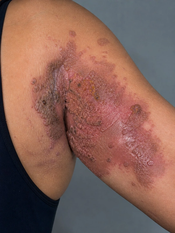 AI illustration
Definition
Pemphigus vegetans is a rare variant of pemphigus vulgaris. It is characterised by vegetating, crusted plaques of excessive granulation tissue, mainly in the large body folds.
Classification
- Neumann type: begins like pemphigus vulgaris with vesicles and bullae; after rupture, hypertrophic granulating erosions develop, later evolving into exuding vegetating masses.
- Hallopeau type: not preceded by bullae; initially pustules that merge after rupture and evolve into vegetating erosions with centrifugal spread.
Occurrence & epidemiology
Pemphigus vegetans accounts for about 1–2% of all pemphigus cases; fewer than 200 cases have been published. In a systematic review (197 patients) the mean age was 52 years with a near-equal sex distribution; the oral mucosa was the first site involved in 44.7%.
Aetiopathogenesis
As in pemphigus vulgaris, IgG autoantibodies against desmosomes disrupt the adhesion between keratinocytes (acantholysis). In the large folds, occlusion, maceration and mixed infections promote exudation and granulation tissue.
Clinical features
- Mainly the axillae, inframammary folds, groins and natal cleft, as well as the face and scalp; the oral mucosa is often involved first.
- In the folds, exuding vegetating plaques; outside the folds they dry out into warty, fissured, painful lesions resembling seborrhoeic keratoses.
Histology
Hyperkeratosis, pseudoepitheliomatous hyperplasia and papillomatosis with suprabasal acantholysis; intraepidermal eosinophilic microabscesses are typical (described in 65% in the review).
Diagnosis
- Histology and direct immunofluorescence (intercellular IgG in 92%, C3 in 53% of cases with data), supplemented by cytology (Tzanck test).
- Serology: indirect immunofluorescence and ELISA for desmoglein antibodies.
- Straightforward when typical pemphigus vulgaris lesions coexist; more difficult with lesions outside the folds.
Differential diagnoses
Keep learning in the app
References (selection)
Cross-references
Note: This page is intended for medical education and does not replace diagnosis or treatment decisions in individual cases.