Eosinophilic fasciitis (Shulman)
- Synonyms
- Shulman, eosinophilic fasciitis, Shulman syndrome
- Specialty
- Dermatology · Inflammatory skin diseases
- Images
- Clinical 2
- In the app
- 2 flashcards
- Last updated
- 10/2026 · Dr. Pascal Bafteh
Contents
Images (2)
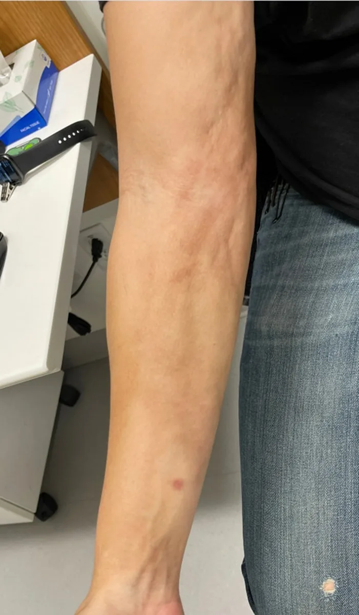
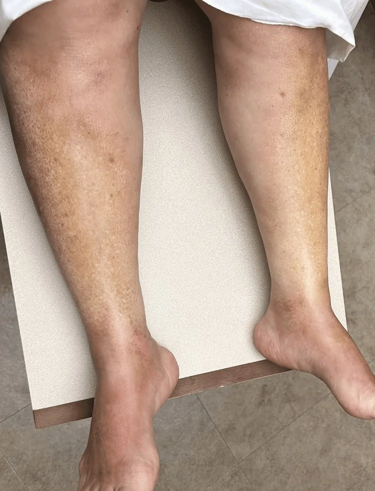
Definition
Eosinophilic fasciitis (Shulman syndrome) is a rare sclerodermiform disease with inflammation and fibrosis of the fascia and subcutaneous tissue, usually with peripheral eosinophilia. The extremities, trunk and neck are mainly affected, usually bilaterally; the digits are characteristically spared. Strenuous physical activity precedes it in about 30%.
- Clinical Features: Characteristic: deep scleroderma-like induration, partly after exertion; initially oedema, later orange-peel skin (mattress phenomenon) and groove sign.
- Sparing Sign: Hands / feet usually free (vs. SSc!), Raynaud's phenomenon typically negative.
- Laboratory: Blood eosinophilia, ↑ESR / CRP, hypergammaglobulinemia, ANA / Scl-70 negative.
- Diagnostics: MRI of the fascia (contrast enhancement), spindle biopsy down to fascia / muscle with evidence of eosinophilic fasciitis + fibrosis.
Differential diagnoses
 Limited systemic sclerosis (CREST syndrome)
 Mycosis fungoides
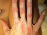 Dermatomyositis
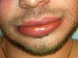 Angioedema
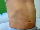 Urticaria
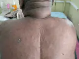 Scleroedema adultorum of Buschke (scleredema)
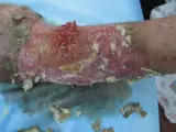 Mucormycosis
Image credits for the thumbnails
- Limited systemic sclerosis (CREST syndrome): James Heilman, MD, “CREST syndrome (calcinosis and sclerodactyly)”, CC BY-SA 4.0, via Wikimedia Commons · CC BY-SA 4.0 · Source
- Mycosis fungoides: Wikindrea, “Mycosis Fungoides Before Sun and Cartamo”, CC BY-SA 4.0, via Wikimedia Commons · CC BY-SA 4.0 · Source
- Dermatomyositis: Mohammad2018, CC BY-SA 4.0 · CC BY-SA 4.0 · Source
- Angioedema: James Heilman, MD, “AngioedemaFra”, CC BY-SA 3.0, via Wikimedia Commons · CC BY-SA 3.0 · Source
- Urticaria: Gzzz, “Urticaria child”, CC BY-SA 4.0, via Wikimedia Commons · CC BY-SA 4.0 · Source
- Scleroedema adultorum of Buschke (scleredema): Ranabahu M, Karunarathna W, Asanthi J et al. (Cureus 2024), CC BY 4.0, via PubMed Central (PMC11178434) · CC BY 4.0 · Source
- Mucormycosis: Ran Yuping et al., “Mucormycosis”, CC BY 3.0, via Wikimedia Commons · CC BY 3.0 · Source · cropped
Treatment principles
- Systemic glucocorticoids are the standard treatment, to which many patients respond well; they are often combined with steroid-sparing immunosuppressants.
- Early treatment and accompanying physiotherapy aim to prevent joint contractures.
- As associations with haematological disorders have been described, their exclusion is part of management.
Specific treatment regimens with drugs and doses are covered in the DermaFuchs app.
Keep learning in the app
References (selection)
Cross-references
Mentioned in
Keep learning
Keep learning with flashcards, exam questions and spot diagnoses in the DermaFuchs app – free, in your browser or as an app.
Related pages: Limited systemic sclerosis (CREST syndrome) · Mycosis fungoides · Dermatomyositis
Note: This page is intended for medical education and does not replace diagnosis or treatment decisions in individual cases.