Diabetic dermopathy (shin spots)
- Synonyms
- Shin spots, Schienbeinflecken, prätibiale pigmentierte Flecken, diabetische Schienbeinflecken, diabetic dermopathy, pigmented pretibial patches
- Specialty
- Dermatology · Metabolic skin disorders
- Images
- Clinical 1
- Last updated
- 10/2026 · Dr. Pascal Bafteh
Contents
Images (1)
Definition
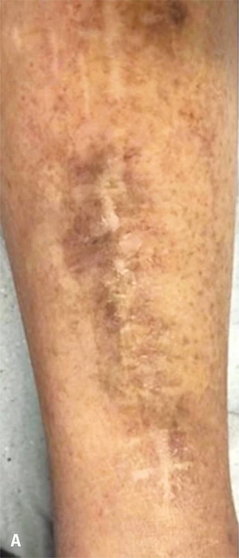
Diabetic dermopathy is one of the most common skin changes in diabetes mellitus. It presents as round to oval, light brown to reddish-brown, slightly depressed (atrophic) patches, typically on the shins ("shin spots").
Occurrence & epidemiology
Up to 30% of people with diabetes are affected, especially older patients or those with diabetes for at least 10–20 years; an association with raised HbA1c has been described. Single lesions also occur without diabetes, particularly after injury; four or more lesions are almost exclusively found in diabetes.
Aetiopathogenesis
The cause is unknown. Dermopathy is associated with the microangiopathic complications retinopathy, neuropathy and nephropathy, and an association with coronary artery disease has also been shown. Because the lesions lie over bony prominences, an exaggerated response to minor trauma is assumed; lesions have been observed after heat, cold or blunt injury.
Clinical features
- Round or oval reddish-brown patches, initially scaly, later flat and depressed.
- Usually on both shins; less often on the front of the thighs, forearm, side of the foot, scalp or trunk.
- The lesions themselves are harmless.
Histology
In an autopsy study (14 biopsies), dermal pigment deposits of haemosiderin and melanin were found in almost all cases, often with increased epidermal melanin; marked vessel wall thickening was seen in only 4 of 14 specimens.
Diagnosis
- Clinical, based on the typical appearance; biopsy is rarely needed.
- Shin spots without known diabetes warrant testing for diabetes mellitus; in known diabetes, dermopathy is regarded as a pointer to microangiopathic complications.
Differential diagnoses
Keep learning in the app
References (selection)
Cross-references
Mentioned in
Note: This page is intended for medical education and does not replace diagnosis or treatment decisions in individual cases.