Keratoacanthoma
Exam relevance: in 3 of more than 50 board exam reports · rank 45
- Synonyms
- KA, Molluscum sebaceum
- Specialty
- Dermatology · Skin tumours
- Images
- Clinical 1 · Histology 3
- In the app
- 8 flashcards
- Last updated
- 10/2026 · Dr. Pascal Bafteh
Contents
Images (4)
 Histology
 Histology
 Histology
Definition
Keratoacanthoma is a rapidly growing, usually solitary epithelial tumour of hair-bearing, sun-exposed skin. It may regress spontaneously with scarring but is often indistinguishable from well-differentiated cutaneous squamous cell carcinoma; many clinicians and pathologists therefore prefer the term "squamous cell carcinoma, keratoacanthoma type".
Occurrence & epidemiology
It mainly affects older, fair-skinned people, men more often than women. Typical sites are the face, back of the hands and forearms. Risk factors are UV exposure and immunosuppression. Multiple keratoacanthomas occur in rare syndromes such as Muir-Torre syndrome and the Ferguson-Smith type.
Clinical features
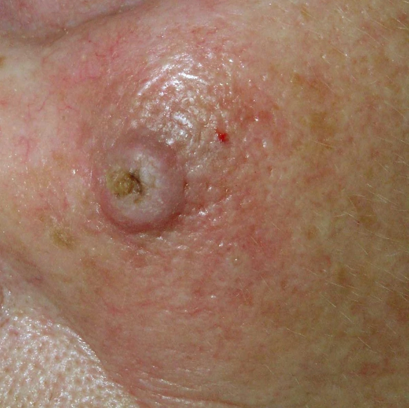
- A dome-shaped, skin-coloured to reddish nodule (usually 1–2 cm) growing within a few weeks, sharply demarcated and firm, with a central keratin plug and an even shoulder; crater-like without the core.
- Typical three-phase course: rapid growth, maturation, then regression leaving a depressed scar; usually 4–6 months from first appearance to complete regression.
- Rapid growth over weeks is the key feature in the history.
Histology
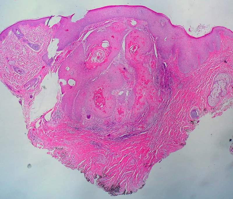
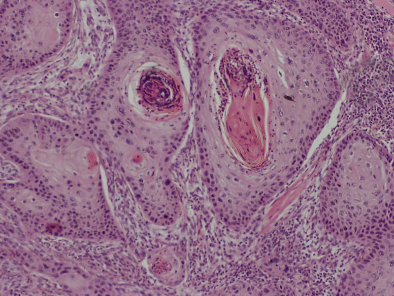
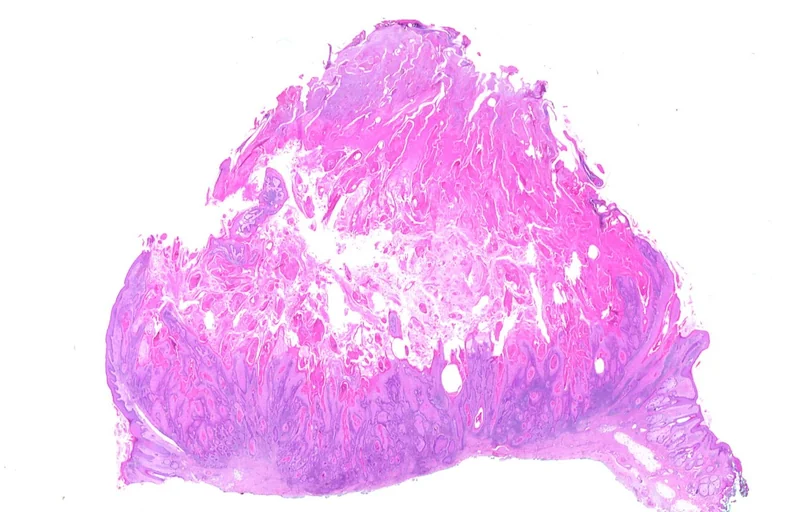
At low power a crateriform architecture with a central keratin plug; enlarged, atypical keratinocytes with eosinophilic cytoplasm that do not extend below the level of the sweat glands. These features often cannot be assessed in partial or shave biopsies.
Diagnosis
- Clinical picture together with growth dynamics.
- Dermoscopy: central keratin plug, ivory-white peripheral zone and symmetrically arranged hairpin vessels; dermoscopy cannot reliably distinguish it from squamous cell carcinoma.
- Histology to confirm the diagnosis and to separate it from squamous cell carcinoma, which can look very similar clinically.
Differential diagnoses
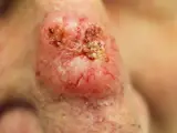 Basal cell carcinoma
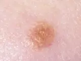 Spitz naevus (spindle and epithelioid cell naevus)
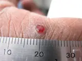 Pyogenic granuloma
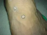 Warts (verrucae vulgares)
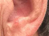 Actinic keratosis
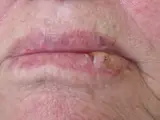 Cutaneous squamous cell carcinoma
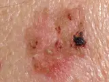 Bowen's disease (squamous cell carcinoma in situ)
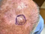 Atypical fibroxanthoma
Image credits for the thumbnails
- Basal cell carcinoma: Bin im Garten, “Basaliom am Nasenrücken 2”, CC BY-SA 3.0, via Wikimedia Commons · CC BY-SA 3.0 · Source
- Spitz naevus (spindle and epithelioid cell naevus): The Armed Forces Institute of Pathology (AFIP), Public domain, via Wikimedia Commons · Public domain · Source
- Pyogenic granuloma: kilbad, “Pyogenic granuloma on a finger-1”, CC BY 3.0, via Wikimedia Commons · CC BY 3.0 · Source
- Warts (verrucae vulgares): Abbassyma (English Wikipedia), “Verruca vulgaris”, Public Domain, via Wikimedia Commons · Public domain · Source
- Actinic keratosis: Future FamDoc, “Actinic keratosis on ear”, CC BY-SA 4.0, via Wikimedia Commons · CC BY-SA 4.0 · Source
- Cutaneous squamous cell carcinoma: Klaus D. Peter, Wiehl, Germany, “Spinaliom3”, CC BY 3.0 DE, via Wikimedia Commons · CC BY 3.0 DE · Source
- Bowen's disease (squamous cell carcinoma in situ): Crop by Mikael Häggström, from original by Klaus D. Peter, Gummersbach, Germany, “Bowen's disease (crop)”, CC BY 3.0 de, via Wikimedia Commons · CC BY 3.0 de · Source
- Atypical fibroxanthoma: Apichoto-Mata I, García-Contreras SJ, Garnica B (Cureus 2026), CC BY 4.0, via PubMed Central (PMC12949712) · CC BY 4.0 · Source
Treatment principles
- Although spontaneous regression occurs, keratoacanthoma is treated like squamous cell carcinoma because the histological distinction is often uncertain.
- The treatment of choice is complete excision with histological examination.
- If surgery is not possible or the site is unfavourable, radiotherapy or intralesional procedures may be considered; follow-up for further epithelial skin tumours is advisable.
Specific treatment regimens with drugs and doses are covered in the DermaFuchs app.
Keep learning in the app
References (selection)
Cross-references
Mentioned in
Keep learning
Keep learning with flashcards, exam questions and spot diagnoses in the DermaFuchs app – free, in your browser or as an app.
Related pages: Basal cell carcinoma · Spitz naevus (spindle and epithelioid cell naevus) · Pyogenic granuloma
Note: This page is intended for medical education and does not replace diagnosis or treatment decisions in individual cases.