Merkel cell carcinoma
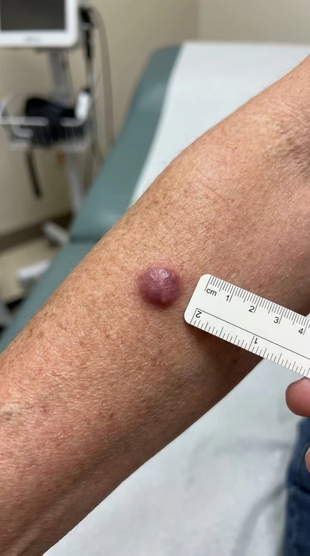
Definition & Epidemiology
- Dermal neuroendocrine tumor with high aggressiveness.
- Cell of origin: Merkel cells / mechanoreceptors — neuronal origin in epidermal basal layer.
- Clinical presentation: bluish-livid nodule — rapidly growing, painless, firm; localization head, neck, extremities (UV-exposed).
AEIOU Features (clinically suspicious)
- Asymptomatic
- Expanding fast (rapid growth)
- Immunosuppression
- Old age (>50 yrs)
- UV-exposed
Pathogenesis & Risk Factors
- Merkel cell polyomavirus (MCPyV): Trigger detectable in 80% — viral oncoproteins LT antigen.
- UV-induced mutations (TP53, RB1) in MCPyV-negative cases — higher mutational burden.
- Immunosuppression: Post-transplant, HIV, hematological diseases, anti-TNF therapy.
- Older age (>70 yrs most common period of manifestation).
Diagnostics
- Punch biopsy (4–6 mm) + Histology + Immunohistochemistry:
- CK20 perinuclear dot-like (Dot-Pattern) — pathognomonic
- CK56, TTF1 typically NEGATIVE (DD to small cell lung carcinoma metastasis, which is TTF1+!)
- Synaptophysin, Chromogranin A (neuroendocrine marker)
- MCPyV-LT-Antigen-IHC (antibody CM2B4) in viral form
- Staging obligatory: PET-CT + SLND — for every MCC, as lymphangiosis cutis is almost obligatory.
- Metastasis pathways: lymphogenous > hematogenous — Lymphangiosis cutis as an early sign, then lung/liver/bone.
Differential diagnoses
- Basal cell carcinoma
- Dermatofibrosarcoma protuberans
- Atypical fibroxanthoma
- Cutaneous angiosarcoma
- Kaposi's sarcoma
- Melanoma
- Pyogenic granuloma
Practise Merkel cell carcinoma in the app
Flashcards with spaced repetition, exam questions and spot-the-diagnosis on this topic – in DermaFuchs, free of charge.
In the DermaFuchs app: 8 flashcards · 6 clinical images
Open in browser Download on the App StoreSources (selection)
More skin tumours
- Basal cell carcinoma
- Melanoma
- Lentigo maligna
- Cutaneous squamous cell carcinoma
- Actinic keratosis
- Actinic cheilitis
- Microcystic adnexal carcinoma
- Dermatofibroma
Note: This page is intended for medical education and does not replace diagnosis or treatment decisions in individual cases. Treatment and follow-up content is available in the app.