Mastocytoma
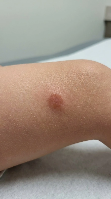
Definition & Classification
- Mastocytoma = benign cutaneous mast cell accumulation in the dermis with local histamine release.
- Forms of cutaneous mastocytosis (CM):
- Solitary mastocytoma (most common form in childhood) — usually single yellow-brown plaque/nodule.
- Urticaria pigmentosa (= maculopapular CM) — multiple red-brown macules/papules on the trunk.
- Diffuse cutaneous mastocytosis (DCM) — rare, entire skin infiltrated, blistering possible.
- Distinction: systemic mastocytosis (SM) involves bone marrow/organs, predominantly in adults with KIT-D816V mutation.
Epidemiology
- Onset usually <2 years of age, sporadic.
- About 90 % of pediatric cutaneous mastocytoses show spontaneous remission by puberty.
Mastocytoma in Children
- Frequency: ~75 % of all cutaneous mastocytoses are pediatric solitary mastocytomas
- First manifestation: typically within the first 2 years of life
- Localization: preferentially trunk, proximal extremities
- Clinical: yellow-brown to reddish plaque, 1–5 cm — Darier's sign positive
- Spontaneous remission: usually by puberty — reassurance of parents important
- Trigger caution (avoid mast cell degranulation): mechanical irritation, heat, aspirin/NSAIDs, opiates, contrast media, alcohol, stress
- ⚠ Anaphylaxis risk with large-area lesions (DCM) → prescribe adrenaline emergency kit
- DD: café-au-lait macule, nevus, juvenile xanthogranuloma
Clinical Features & Darier's Sign
- Typical lesion: yellow-brown plaque or papule, single, often 1–5 cm, trunk/extremities.
- Darier's sign positive: mechanical stroking → urticarial wheal + erythema, possibly blister due to mast cell degranulation.
- Accompanying symptoms (with histamine release): flushing, pruritus, possibly blistering.
- Caution: extensive DCM → anaphylaxis risk with triggers.
Diagnostics
- Clinical: inspection + Darier's sign (rub gently, not vigorously — avoid anaphylaxis risk!).
- Biopsy if suspected: histology + toluidine blue staining (metachromatic) or CD117/tryptase IHC.
- Lab: serum tryptase (baseline) to rule out SM — values > 20 ng/ml suggestive of systemic mastocytosis (additionally bone marrow biopsy + KIT-D816V mutation analysis).
- DD: café-au-lait macule, juvenile xanthogranuloma, melanocytic nevus.
Differential diagnoses
- Haemangioma
- Dermatofibroma
- Melanoma
- Cutaneous angiosarcoma
- Kimura disease
- Urticaria
- Mastocytosis (urticaria pigmentosa)
Practise Mastocytoma in the app
Flashcards with spaced repetition, exam questions and spot-the-diagnosis on this topic – in DermaFuchs, free of charge.
In the DermaFuchs app: 2 flashcards · 2 clinical images
Open in browser Download on the App StoreSources (selection)
- Valent 2022 WHO mastocytosis classification
- Hartmann 2016 mastocytosis ECNM consensus
- Valent 2021 mastocytosis review NEJM
More skin tumours
- Basal cell carcinoma
- Melanoma
- Lentigo maligna
- Cutaneous squamous cell carcinoma
- Actinic keratosis
- Actinic cheilitis
- Merkel cell carcinoma
- Microcystic adnexal carcinoma
Note: This page is intended for medical education and does not replace diagnosis or treatment decisions in individual cases. Treatment and follow-up content is available in the app.