Spitz naevus (spindle and epithelioid cell naevus)

Synonyms
Spitz-Nävus, Spitz-Naevus, Spindelzellnävus, Epitheloidzellnävus, Reed-Nävus, pigmentierter Spindelzelltumor Reed, Spitz nevus, Reed naevus
Specialty
Dermatology · Skin tumours
Images
Clinical 1 · Dermoscopy 1 · Histology 4
In the app
1 flashcards
Last updated
10/2026 · Dr. Pascal Bafteh
Contents
  1. Images (6)
  2. Definition
  3. Occurrence & epidemiology
  4. Aetiopathogenesis
  5. Clinical features
  6. Histology
  7. Diagnosis
  8. Differential diagnoses
  9. Keep learning in the app
  10. References (selection)
  11. Cross-references

Images (6)

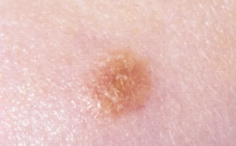
Spitz naevus (spindle and epithelioid cell naevus) – clinical photo: small, symmetrical, uniformly pink-tan papule
small, symmetrical, uniformly pink-tan papuleThe Armed Forces Institute of Pathology (AFIP), Public domain, via Wikimedia Commons · Public domain · Source
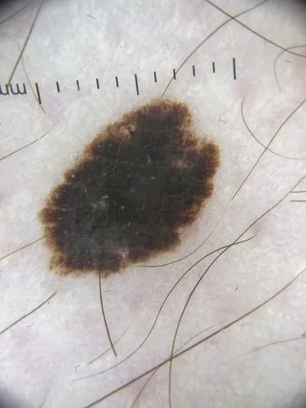
Spitz naevus (spindle and epithelioid cell naevus) – dermoscopy: starburst pattern with a black centre and a closed rim of streaksDermoscopy
dermoscopy: starburst pattern with a black centre and a closed rim of streaks. (Photo: Pascal Bafteh)Photo: KLINIKFUCHS · published with patient consent
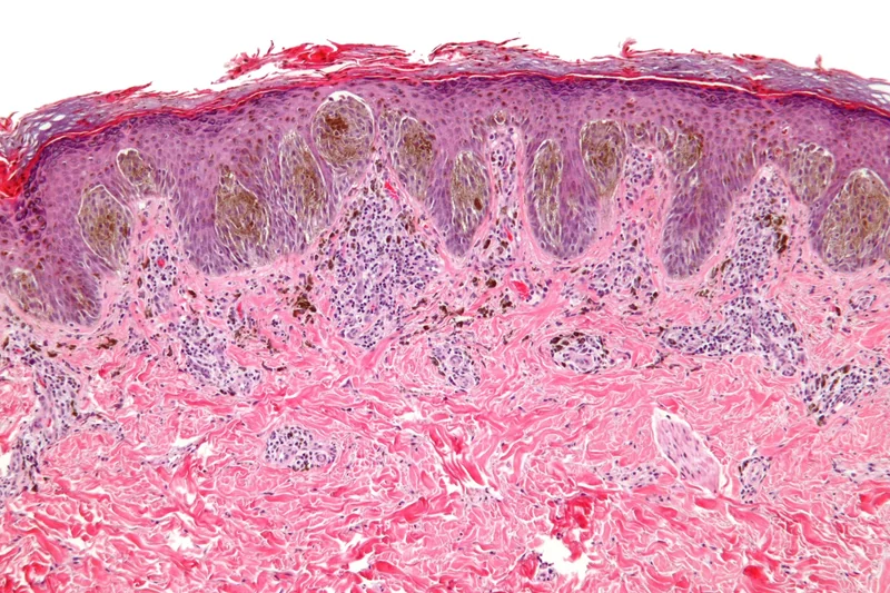
Spitz naevus (spindle and epithelioid cell naevus) – histology (H&E), overview: a symmetric, superficially confined junctional melanocytic proliferation with heavily pigmented nests at the tips of elongated rete ridges, a band-like …Histology
histology (H&E), overview: a symmetric, superficially confined junctional melanocytic proliferation with heavily pigmented nests at the tips of elongated rete ridges, a band-like …Nephron, „Spitz nevus - intermed mag", CC BY-SA 3.0, via Wikimedia Commons · CC BY-SA 3.0 · Source
Spitz naevus (spindle and epithelioid cell naevus) – histologyHistology
Spitz naevus (spindle and epithelioid cell naevus) – histologyLWozniak&KWZielinski, „SkinTumors-446", CC BY-SA 3.0, via Wikimedia Commons · CC BY-SA 3.0 · Source
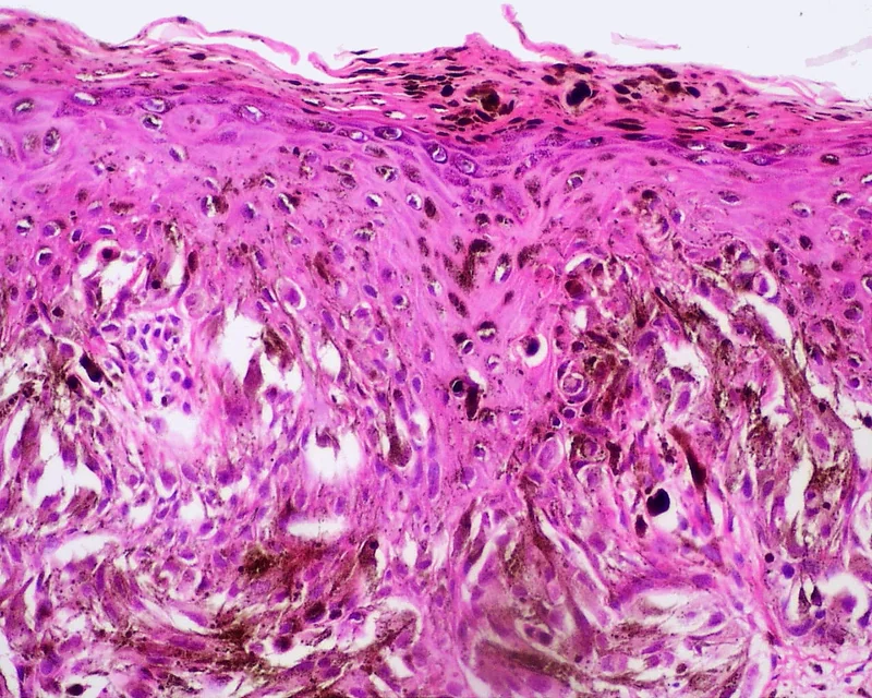
Spitz naevus (spindle and epithelioid cell naevus) – histology (H&E), heavily overstained field: abundant melanin in the upper dermis (melanophages) and pigmented cells reaching the upper epidermal layers; individual junctional nests …Histology
histology (H&E), heavily overstained field: abundant melanin in the upper dermis (melanophages) and pigmented cells reaching the upper epidermal layers; individual junctional nests …LWozniak&KWZielinski, „Skin Tumors-263", CC BY-SA 3.0, via Wikimedia Commons · CC BY-SA 3.0 · Source
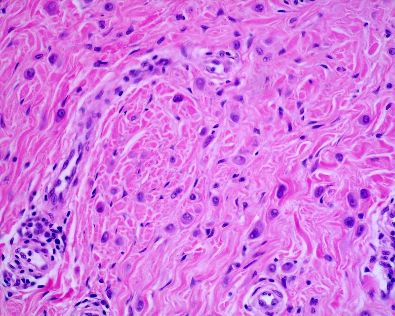
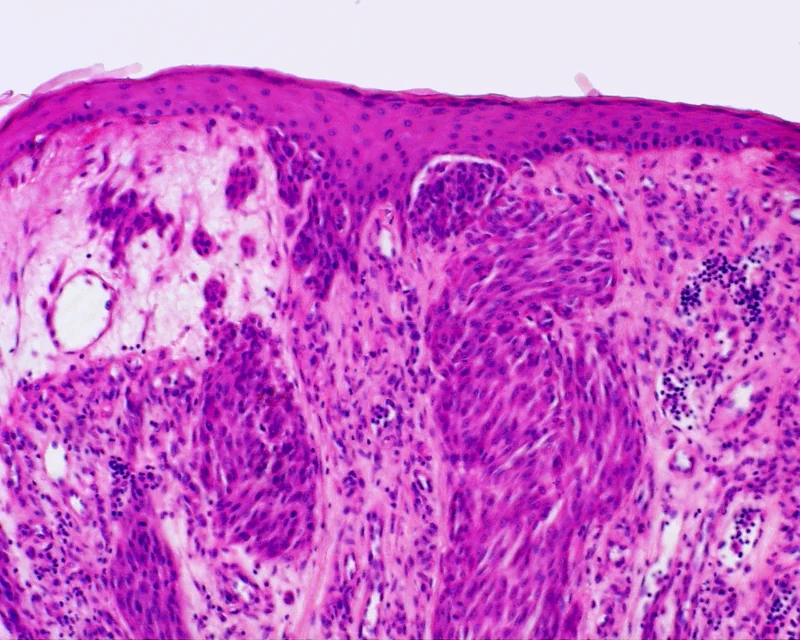
Spitz naevus (spindle and epithelioid cell naevus) – histology (H&E): nests of large spindled and epithelioid melanocytesHistology
histology (H&E): nests of large spindled and epithelioid melanocytesLWozniak&KWZielinski, „SkinTumors-214", CC BY-SA 3.0, via Wikimedia Commons · CC BY-SA 3.0 · Source
1 / 6

Definition

A Spitz naevus is an uncommon, benign melanocytic naevus composed of epithelioid and/or spindle-shaped melanocytes. Clinically and histologically it may resemble melanoma. It belongs to the spectrum of Spitz tumours, which comprises Spitz naevus, Spitz melanocytoma (intermediate) and rare Spitz melanoma.

Occurrence & epidemiology

Spitz naevi occur mainly in children; 70% are diagnosed in the first 20 years of life. They also arise in adults and are most frequent in fair skin.

Aetiopathogenesis

A distinct molecular pathway is characteristic, mainly kinase fusions or HRAS aberrations; it separates bona fide Spitz neoplasms from Spitz-like naevi and melanomas with conventional driver mutations.

Clinical features

  • Classic: dome-shaped red to reddish-brown papule.
  • Pigmented: tan to brown papule or nodule.
  • Pigmented spindle cell tumour of Reed: bluish-black papule.
  • Size from a few millimetres to 1–2 cm, mainly on the face and limbs, usually solitary.
  • Rapid growth over a few months is typical, often followed by years without change; spontaneous regression is possible.

Histology

Usually a symmetrical compound naevus made of nests of characteristic epithelioid cells (classic and pigmented types) or spindle cells (Reed); both cell types can coexist. Distinguishing atypical Spitz tumours from melanoma is often difficult histologically.

Diagnosis

  • Dermoscopy: classic type with evenly distributed dotted or rounded vessels; pigmented type with a starburst or globular pattern, later homogeneous; Reed type with a deeply pigmented starburst pattern with pseudopods. No pattern reliably excludes melanoma.
  • Histological confirmation, particularly in older children and adults; molecular studies help in unclear cases.

Differential diagnoses

Keep learning in the app

In the DermaFuchs app you can learn Spitz naevus (spindle and epithelioid cell naevus) with flashcards, exam questions and spot-the-diagnosis – free, in your browser or as an app.

Test yourself: Spot-diagnosis quiz with real cases →

In the app: 1 flashcards

Open in browser  Download on the App Store

References (selection)

  1. DermNet: Spitz naevus
  2. PubMed: Spitz melanocytic tumours - a review (PMID 34958498)
  3. PubMed: Spitz nevus, Spitz tumor, and spitzoid melanoma: a comprehensive clinicopathologic overview (PMID 24075547)

Cross-references

Note: This page is intended for medical education and does not replace diagnosis or treatment decisions in individual cases.