Warts (verrucae vulgares)
- Synonyms
- Warze, Verruca vulgaris, Dornwarze, Fußwarze, Plantarwarze, flache Warzen, Verrucae planae, HPV, warts, verruca, plantar wart, common wart, flat warts
- Specialty
- Dermatology · Viral infections
- Images
- Clinical 8 · Histology 4
- In the app
- 5 flashcards
- Last updated
- 10/2026 · Dr. Pascal Bafteh
Contents
Images (12)
 Histology
 Histology
 Histology
 Histology
Definition
Warts (verrucae) are benign growths of skin and mucosa caused by human papillomaviruses (HPV). Appearance and course differ according to HPV type and site; anogenital warts are considered separately.
Classification
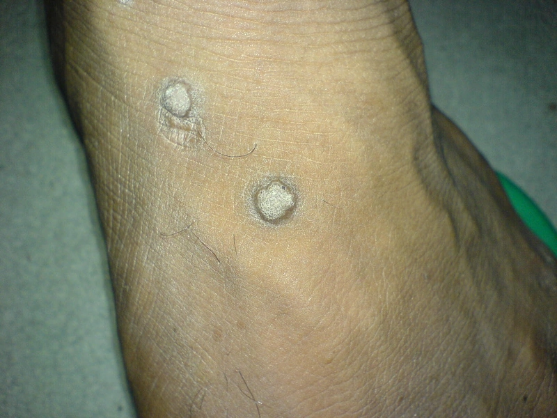
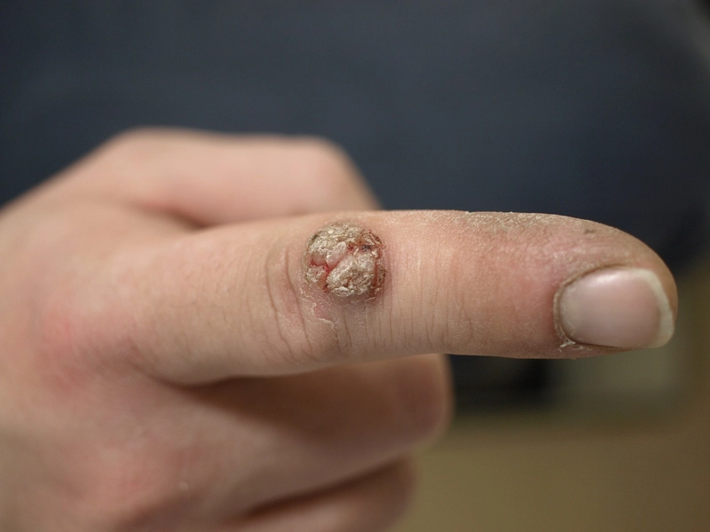
- Common warts (verrucae vulgares): cauliflower-like, 1 mm to over 1 cm, mainly on the knees, backs of fingers and toes and periungually.
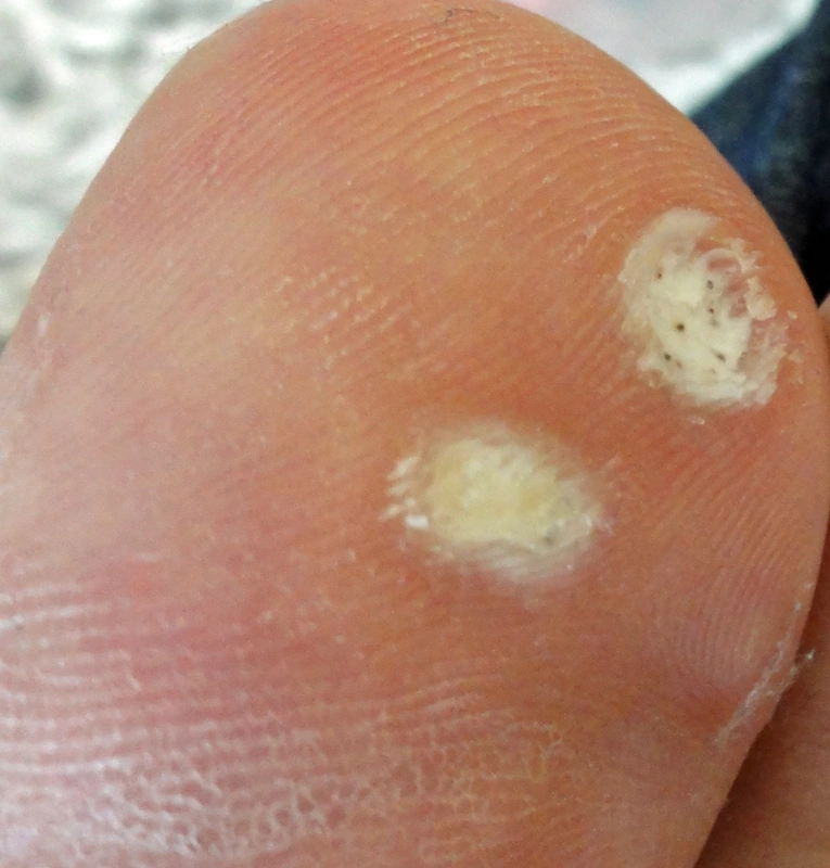
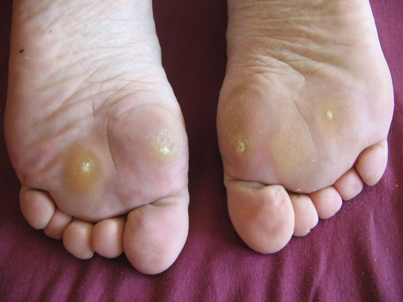
- Plantar warts: endophytic growth into the depth, painful when walking; mosaic warts as flat clusters.
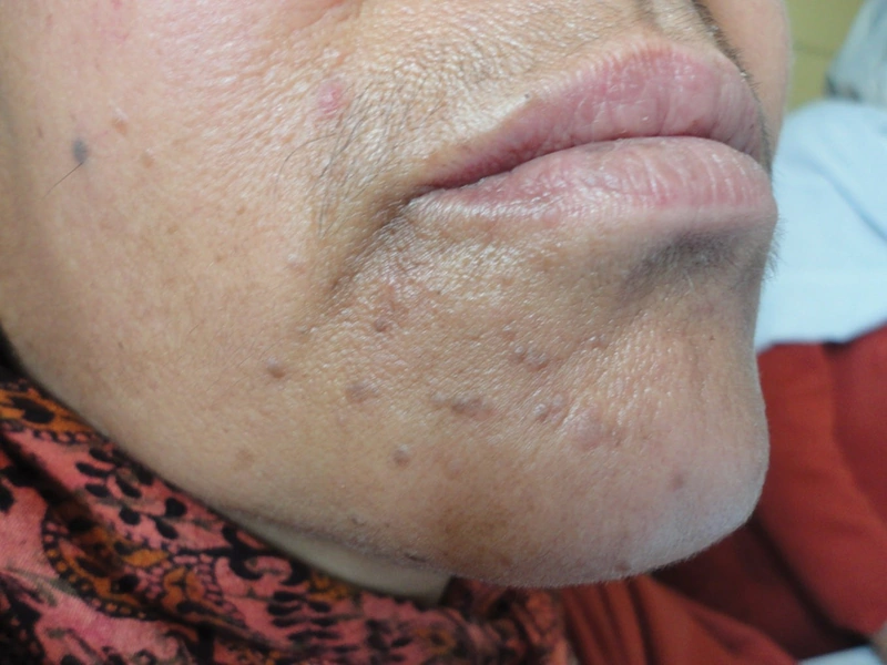
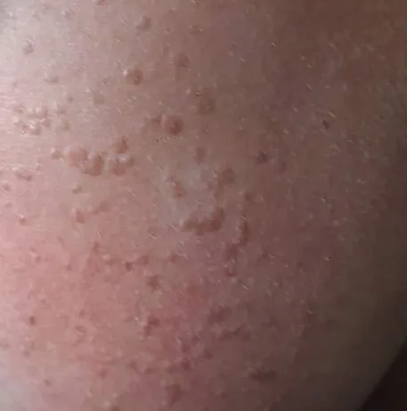
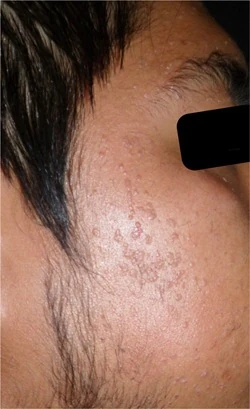
- Plane (flat) warts (verrucae planae): usually multiple small, flat, skin-coloured papules on the face, hands and shins, often spread by shaving; mostly HPV 3 and 10.
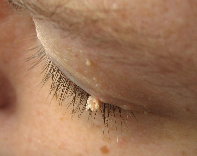
- Filiform warts: thread-like, mainly on the face.
Occurrence & epidemiology
Warts are very common, especially in school-age children and adolescents. Transmission is by direct skin contact or autoinoculation (e.g. by scratching; warts along scratch lines); the incubation period can be up to 12 months; an impaired skin barrier (dermatitis) favours infection. In drug-induced immunosuppression or HIV infection warts are more common and more likely to recur. In children 50% clear within six months and 90% within two years; in adults warts are more persistent.
Clinical features
- Skin-coloured to greyish-brown, firm papules with a rough, fissured surface (papillomatosis).
- Red or black dots within the wart correspond to papillary capillaries and are typical; paring causes pinpoint bleeding.
- Plantar warts interrupt the skin lines and are painful on lateral pressure.
Histology
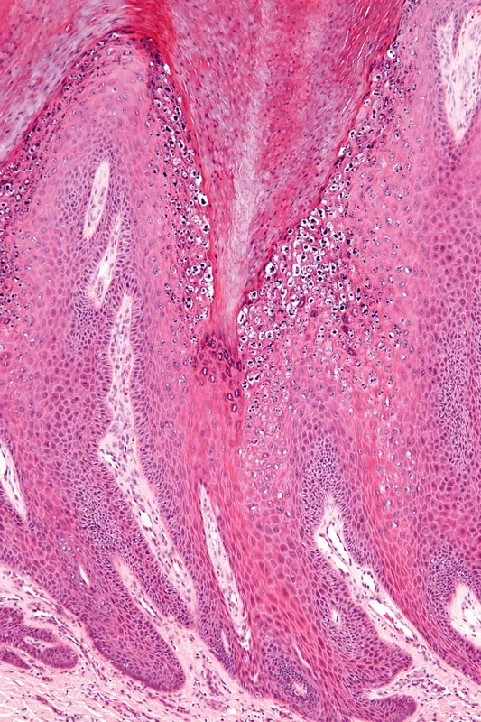
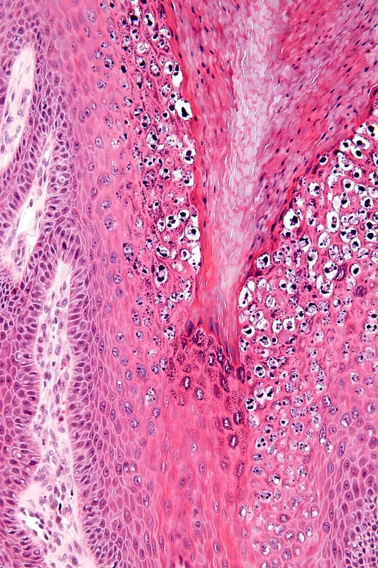
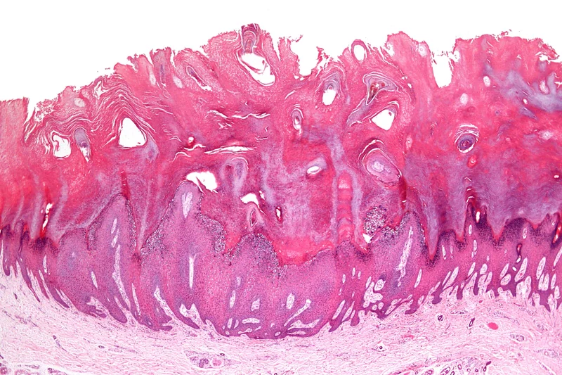
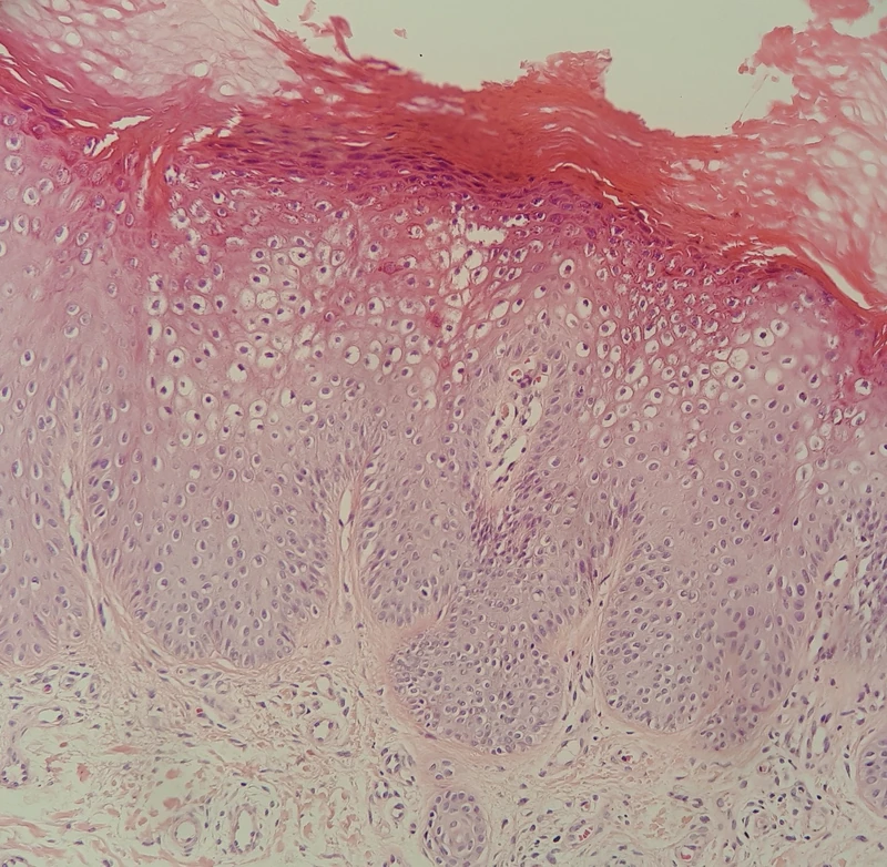
Markedly papillomatous epidermis with hypergranulosis and overlying tiers of parakeratosis; koilocytes (vacuolated keratinocytes with a small shrunken nucleus and perinuclear halo) in the upper epidermis, sometimes large pink inclusions or small basophilic granules.
Diagnosis
- Usually a spot diagnosis; dermoscopy shows red to black dots (capillaries) within papillomatous structures.
- For plantar warts, distinction from corn and callus: these are always at pressure sites, lack papillary capillaries and are more tender with direct pressure.
- Further differentials are seborrhoeic keratosis and squamous cell carcinoma; biopsy is needed when squamous cell carcinoma cannot be excluded clinically (e.g. in organ transplant recipients).
Differential diagnoses
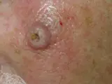 Keratoacanthoma
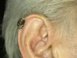 Cutaneous horn (cornu cutaneum)
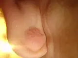 Epidermoid cyst
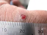 Pyogenic granuloma
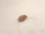 Seborrhoeic keratosis
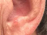 Actinic keratosis
- Verruca plana
Image credits for the thumbnails
- Keratoacanthoma: Klaus D. Peter, Wiehl, Germany, “Spinaliom”, CC BY 3.0 DE, via Wikimedia Commons · CC BY 3.0 DE · Source
- Cutaneous horn (cornu cutaneum): Klaus D. Peter, Wiehl, Germany, “Cornu cutaneum2”, CC BY 3.0 DE, via Wikimedia Commons · CC BY 3.0 DE · Source
- Epidermoid cyst: unknown (Commons user), “Epidermal Cyst ear”, CC BY-SA 3.0, via Wikimedia Commons · CC BY-SA 3.0 · Source
- Pyogenic granuloma: kilbad, “Pyogenic granuloma on a finger-1”, CC BY 3.0, via Wikimedia Commons · CC BY 3.0 · Source
- Seborrhoeic keratosis: Assafn, “Seborrheic keratosis closup”, CC BY-SA 4.0, via Wikimedia Commons · CC BY-SA 4.0 · Source
- Actinic keratosis: Future FamDoc, “Actinic keratosis on ear”, CC BY-SA 4.0, via Wikimedia Commons · CC BY-SA 4.0 · Source
Treatment principles
- Common warts often regress spontaneously, particularly in children, so watchful waiting is a reasonable option.
- Treatment is stepwise, with keratolytic preparations and mechanical debridement, cryotherapy and, for recalcitrant warts, laser or immunomodulatory methods.
- Surgical excision is avoided because of scarring and the risk of recurrence; in immunosuppression, warts are often resistant to treatment.
Specific treatment regimens with drugs and doses are covered in the DermaFuchs app.
Keep learning in the app
References (selection)
Cross-references
Mentioned in
Keep learning
Keep learning with flashcards, exam questions and spot diagnoses in the DermaFuchs app – free, in your browser or as an app.
Related pages: Keratoacanthoma · Cutaneous horn (cornu cutaneum) · Epidermoid cyst
Note: This page is intended for medical education and does not replace diagnosis or treatment decisions in individual cases.