Congenital haemangioma (RICH, PICH, NICH)
- Synonyms
- NICH, RICH, PICH, nicht involutierendes kongenitales Hämangiom, rasch involutierendes kongenitales Hämangiom, partiell involutierendes kongenitales Hämangiom, congenital hemangioma, noninvoluting congenital hemangioma, rapidly involuting congenital hemangioma
- Specialty
- Dermatology · Skin tumours
- Images
- Clinical 1
- In the app
- 2 flashcards
- Last updated
- 10/2026 · Dr. Pascal Bafteh
Contents
Images (1)
Definition
Congenital haemangiomas are rare, usually solitary vascular tumours that grow in utero, are fully formed at birth and do not proliferate further afterwards. Unlike infantile haemangioma, their endothelial cells do not express GLUT1.
Classification
- RICH (rapidly involuting): regression in the first months of life – in one series, in all 24 cases left to their natural course by 14 months of age; atrophic skin or excess skin remains.
- PICH (partially involuting): rapid regression during the first 12–30 months of life, after which a NICH-like lesion persists.
- NICH (non-involuting): non-progressive, persists permanently.
Aetiopathogenesis
RICH and NICH carry the same somatic activating mutations in GNAQ or GNA11; their different course is therefore due to other factors.
Clinical features
- RICH and NICH are similar in appearance, location and size.
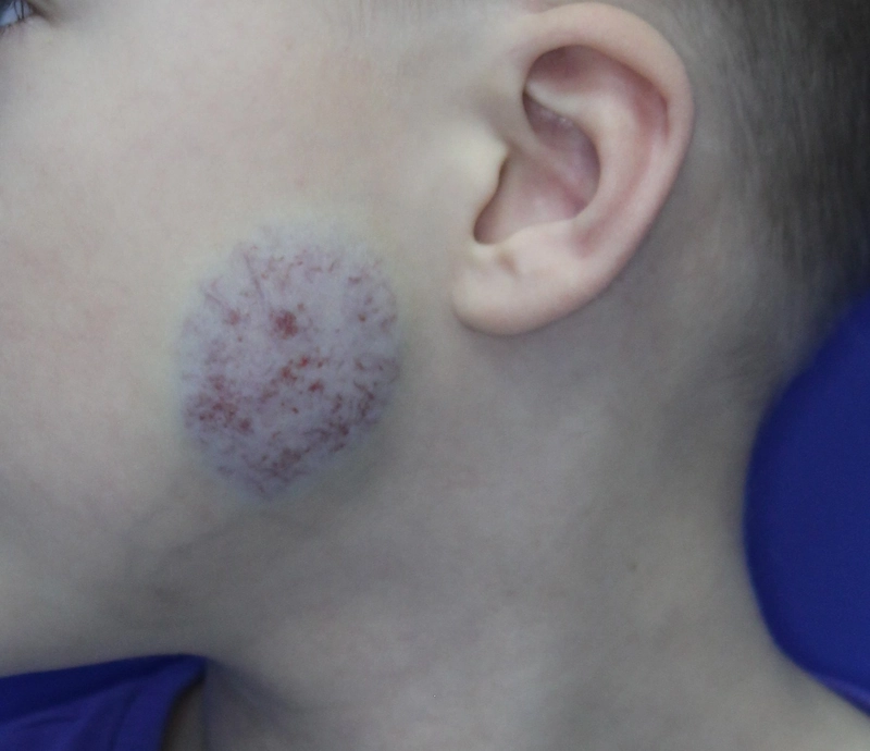
- Most common forms (series of 31 infants): raised violaceous tumour with ectatic veins; raised greyish tumour with fine telangiectases and a pale halo; flat infiltrative tumour with violaceous overlying skin.
- RICH may ulcerate or bleed early.
- NICH (series of 30 cases): mainly trunk and legs, girls more often; 13 of 30 reported pain.
Histology
Capillary lobules in dense fibrotic stroma with haemosiderin, focal thrombosis and thin-walled vessels; GLUT1-negative (in a systematic review all 16 congenital haemangiomas were negative, whereas 95% of infantile haemangiomas were positive).
Diagnosis
- Decisive: fully formed at birth, without a postnatal growth phase – infantile haemangioma, by contrast, appears perinatally and grows rapidly in the first year of life.
- Doppler ultrasound: high blood flow (described for NICH).
- GLUT1 staining if histology is unclear: a negative result makes infantile haemangioma unlikely.
Differential diagnoses
Keep learning in the app
References (selection)
- Congenital hemangiomas
- Congenital hemangioma: evidence of accelerated involution
- Partially involuting congenital hemangiomas: a report of 8 cases and review of the literature
- Characteristics of noninvoluting congenital hemangioma: a retrospective review
- Congenital nonprogressive hemangioma: a distinct clinicopathologic entity unlike infantile hemangioma
- The utility of GLUT1 as a diagnostic marker in cutaneous vascular anomalies: A review of literature and recommendations for daily practice
- Somatic Activating Mutations in GNAQ and GNA11 Are Associated with Congenital Hemangioma
- Rapidly involuting congenital hemangioma: clinical and histopathologic features
Cross-references
Note: This page is intended for medical education and does not replace diagnosis or treatment decisions in individual cases.