Cutaneous pseudolymphoma
Exam relevance: in 1 of more than 50 board exam reports · rank 93
- Synonyms
- Lymphadenosis cutis benigna, reaktive lymphoide Hyperplasie, pseudolymphoma
- Specialty
- Dermatology · Skin tumours
- Images
- Clinical 2 · Histology 1
- In the app
- 5 flashcards
- Last updated
- 10/2026 · Dr. Pascal Bafteh
Contents
Images (3)
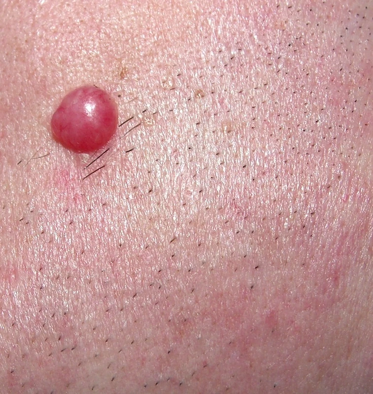
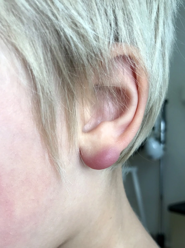
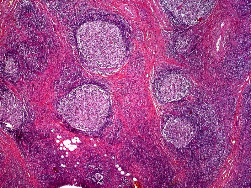 Histology
Definition
- Definition: benign, polyclonal lymphoreticular proliferations, difficult to distinguish from cutaneous lymphomas.
- Histological differentiation from malignant lymphoma: Polyclonality (no light-chain restriction), usually more T than B cells; growth pattern ("top-/bottom-heavy") and reactive follicles alone are not diagnostic.
Classification
Classification by Etiology
Virus-induced Pseudolymphomas
- HSV infection as a trigger for a pseudolymphomatous reaction (Leinweber et al. 2006).
- EBV, Molluscum, HHV infections also described.
Unexplained Etiology
- Lymphomatoid Papulosis (LyP): CD30+ T-cell lymphoma with recurrent lesions that heal spontaneously. Histological types A–E plus a type with 6p25.3 (DUSP22) rearrangement.
- Actinic Reticuloid (CAD): chronic dermatitis primarily in older men, histology MF-like.
Differential diagnoses
 Dermatofibroma
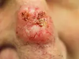 Basal cell carcinoma
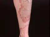 Granuloma annulare
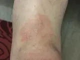 Mycosis fungoides
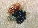 Melanoma
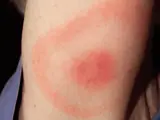 Erythema migrans (Lyme disease)
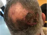 Cutaneous angiosarcoma
- Borrelia pseudolymphoma
Image credits for the thumbnails
- Dermatofibroma: Jmarchn, “Pigmented and tenderness dermatofibroma”, CC BY-SA 3.0, via Wikimedia Commons · CC BY-SA 3.0 · Source
- Basal cell carcinoma: Bin im Garten, “Basaliom am Nasenrücken 2”, CC BY-SA 3.0, via Wikimedia Commons · CC BY-SA 3.0 · Source
- Granuloma annulare: Kreuter et al., “Generalized granuloma annulare”, CC BY-SA 2.0, via Wikimedia Commons · CC BY-SA 2.0 · Source
- Mycosis fungoides: Wikindrea, “Mycosis Fungoides Before Sun and Cartamo”, CC BY-SA 4.0, via Wikimedia Commons · CC BY-SA 4.0 · Source
- Melanoma: National Cancer Institute (unknown photographer), “Melanoma (4)”, Public Domain, via Wikimedia Commons · Public domain · Source
- Erythema migrans (Lyme disease): James Gathany (CDC), “Erythema migrans - erythematous rash in Lyme disease - PHIL 9875”, Public Domain, via Wikimedia Commons · Public domain · Source · CDC PHIL #9875
- Cutaneous angiosarcoma: Messias H, Martins M, Zagalo C et al. (Cancer Rep (Hoboken) 2022), CC BY 4.0, via PubMed Central (PMC9575495) · CC BY 4.0 · Source
Treatment principles
- The key step is identifying and eliminating the trigger, for example stopping a drug, avoiding a contact allergen or treating Lyme borreliosis with antibiotics.
- Topical or intralesional glucocorticoids are used locally.
- For persistent lesions, phototherapy, local radiotherapy or excision may be considered; because of the differential diagnosis of cutaneous lymphoma, follow-up and, if necessary, repeat biopsies are indicated.
Specific treatment regimens with drugs and doses are covered in the DermaFuchs app.
Keep learning in the app
References (selection)
Cross-references
Keep learning
Keep learning with flashcards, exam questions and spot diagnoses in the DermaFuchs app – free, in your browser or as an app.
Related pages: Dermatofibroma · Basal cell carcinoma · Granuloma annulare
Note: This page is intended for medical education and does not replace diagnosis or treatment decisions in individual cases.