Cutaneous squamous cell carcinoma
Exam relevance: in 6 of more than 50 board exam reports · rank 24
- Synonyms
- Spinaliom, Stachelzellkarzinom, cSCC, squamous cell carcinoma, SCC, non-melanoma skin cancer
- Specialty
- Dermatology · Skin tumours
- Images
- Clinical 3 · Histology 2 · Dermoscopy 1
- In the app
- 26 flashcards
- Last updated
- 10/2026 · Dr. Pascal Bafteh
Contents
Images (6)
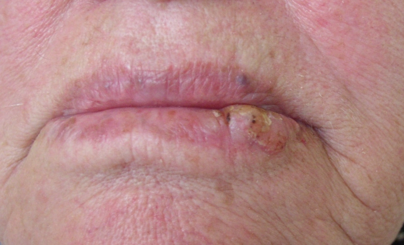
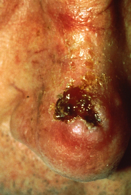
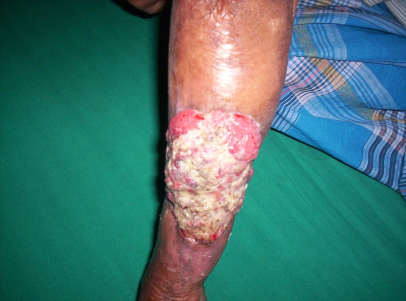
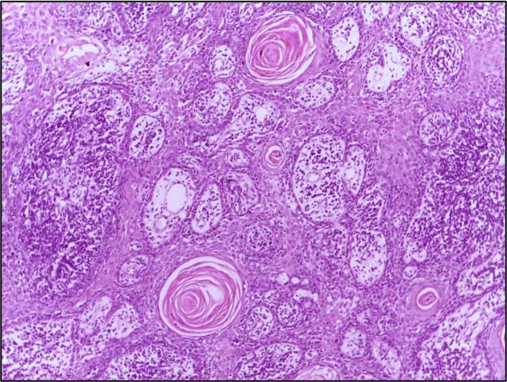 Histology
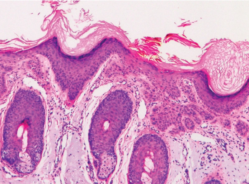 Histology
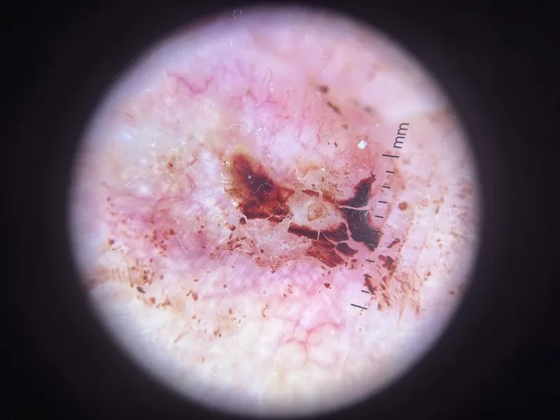 Dermoscopy
Definition
Definition & Origin
- Malignant epithelial tumor originating from the keratinocytes of the epidermis.
- Most frequent mutations: Typical mutations are TP53 + NOTCH with RAS pathway activation.
- Pathogenesis: chronic UV exposure → cumulative DNA damage → Actinic Keratosis → carcinoma in situ (Bowen's disease) → invasive cSCC.
Aetiopathogenesis
Risk Factors — High-Risk Criteria (DDG S3 Guideline 032-022OL)
Tumor-related
- A cSCC is already high-risk if a single criterion is met, e.g. TD > 6 mm or Ø > 2 cm; likewise desmoplasia or perineural invasion.
- Location: High-risk areas are lower lip + ear with high metastatic risk.
- High-risk criteria at a glance: the triad regularly asked for is tumour thickness / vertical depth of invasion, horizontal diameter and degree of differentiation; added to these are desmoplasia, perineural invasion, site, recurrence and immunosuppression.
| Criterion | High risk from |
|---|---|
| Vertical tumour thickness / depth of invasion | > 6 mm, or invasion beyond the subcutaneous fat (muscle, cartilage, bone) |
| Horizontal diameter | > 2 cm |
| Degree of differentiation (grading) | G3/G4 — poorly differentiated to undifferentiated |
| Desmoplasia | present — markedly increased risk of local recurrence and metastasis |
| Perineural invasion (PNI) | present, especially with nerve calibre ≥ 0.1 mm |
| Site | ear and lip, other high-risk facial areas |
| Host factor | immunosuppression (organ transplantation, haematological neoplasia, HIV) |
Patient-related
- Immunosuppression: History of organ transplantation (OTX), HIV, hematological diseases — after organ transplantation the risk is increased 65–250-fold.
- Genodermatoses: Xeroderma pigmentosum, EB dystrophica.
- Muir-Torre Syndrome: Characteristic are sebaceous gland tumors + KA + visceral tumors as a variant of Lynch syndrome (MMR mutation).
Exam essentials
- Key point: cutaneous squamous cell carcinoma usually arises on chronically sun-damaged skin, often from actinic keratoses; the in-situ stage is Bowen's disease.
- Main risk factors: cumulative UV exposure, fair skin type, older age and immunosuppression – especially after organ transplantation.
- Clinical picture: indurated, often hyperkeratotic nodule or plaque that grows, bleeds or ulcerates; mainly face, ears, lower lip, backs of the hands and scalp.
- Diagnosis is histological: atypical keratinocytes infiltrating the dermis, often with keratin pearls.
- Key differential diagnoses: keratoacanthoma (rapid growth, central keratin crater), basal cell carcinoma, hypertrophic actinic keratosis.
Differential diagnoses
 Basal cell carcinoma
 Keratoacanthoma
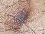 Dermatofibroma
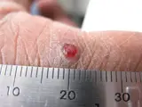 Pyogenic granuloma
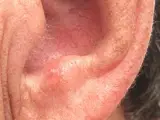 Actinic keratosis
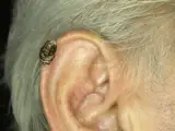 Cutaneous horn (cornu cutaneum)
Image credits for the thumbnails
- Basal cell carcinoma: Bin im Garten, “Basaliom am Nasenrücken 2”, CC BY-SA 3.0, via Wikimedia Commons · CC BY-SA 3.0 · Source
- Keratoacanthoma: Klaus D. Peter, Wiehl, Germany, “Spinaliom”, CC BY 3.0 DE, via Wikimedia Commons · CC BY 3.0 DE · Source
- Dermatofibroma: Jmarchn, “Pigmented and tenderness dermatofibroma”, CC BY-SA 3.0, via Wikimedia Commons · CC BY-SA 3.0 · Source
- Pyogenic granuloma: kilbad, “Pyogenic granuloma on a finger-1”, CC BY 3.0, via Wikimedia Commons · CC BY 3.0 · Source
- Actinic keratosis: Future FamDoc, “Actinic keratosis on ear”, CC BY-SA 4.0, via Wikimedia Commons · CC BY-SA 4.0 · Source
- Cutaneous horn (cornu cutaneum): Klaus D. Peter, Wiehl, Germany, “Cornu cutaneum2”, CC BY 3.0 DE, via Wikimedia Commons · CC BY 3.0 DE · Source
Related conditions: how to tell them apart
- Actinic keratosis – the most common precursor on sun-damaged skin
- Bowen's disease (squamous cell carcinoma in situ) – squamous cell carcinoma in situ
- Keratoacanthoma – rapidly growing nodule with keratin crater – often hard to separate histologically
- Basal cell carcinoma – most common skin cancer, pearly border, rarely metastasises
- Favre-Racouchot disease – another sign of chronic UV damage on the face
Treatment principles
- The treatment of choice is complete excision with histological margin control, preferably micrographically controlled for high-risk tumours and in functionally sensitive sites.
- Radiotherapy is an option for inoperable tumours or as an adjunct after R1 resection; advanced or metastatic tumours are discussed at a multidisciplinary tumour board and treated systemically, mainly with PD-1 inhibitors.
- Strict sun protection and risk-adapted follow-up are part of management.
Guideline: AWMF guideline register (no. 032-022OL)
Specific treatment regimens with drugs and doses are covered in the DermaFuchs app.
Keep learning in the app
References (selection)
Cross-references
Mentioned in
Keep learning: test yourself
One question from the DermaFuchs app on Cutaneous squamous cell carcinoma – tap an answer.
Question 1 of 1
Which mutations are most frequently found in cutaneous squamous cell carcinoma (cSCC)?
Answer
Correct: TP53 + NOTCH. TP53 and NOTCH mutations are the most frequent genetic alterations in cutaneous squamous cell carcinoma.
More questions like these – with explanations and spaced repetition – in the app.
Note: This page is intended for medical education and does not replace diagnosis or treatment decisions in individual cases.