Lentigo maligna
Exam relevance: in 1 of more than 50 board exam reports · rank 93
- Synonyms
- Lentigo-maligna-Melanom, LMM, Hutchinson's freckle
- Specialty
- Dermatology · Skin tumours
- Images
- Clinical 2 · Histology 4
- In the app
- 2 flashcards
- Last updated
- 10/2026 · Dr. Pascal Bafteh
Contents
Images (6)
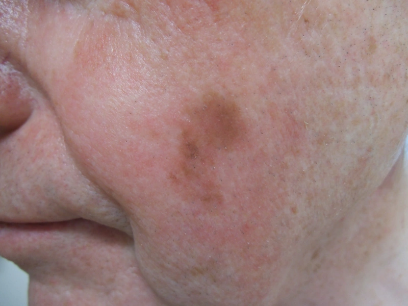
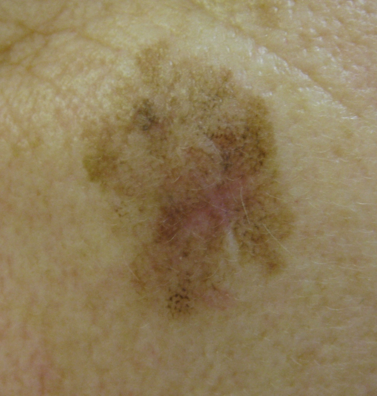
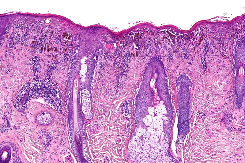 Histology
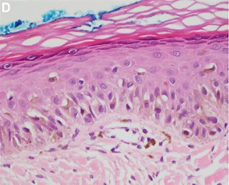 Histology
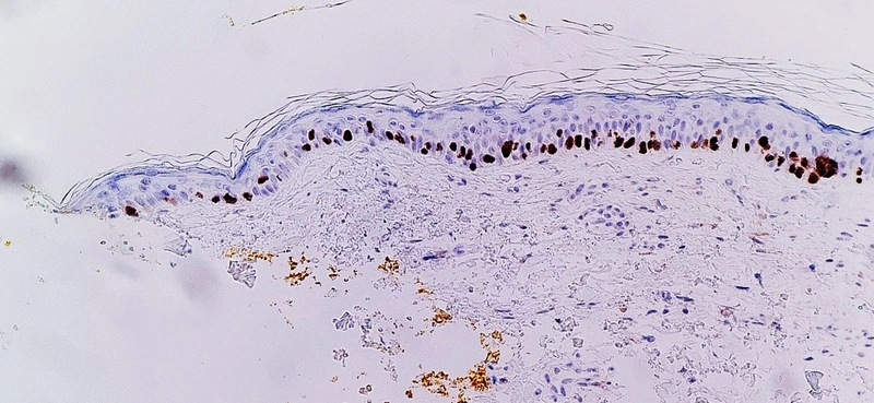 Histology
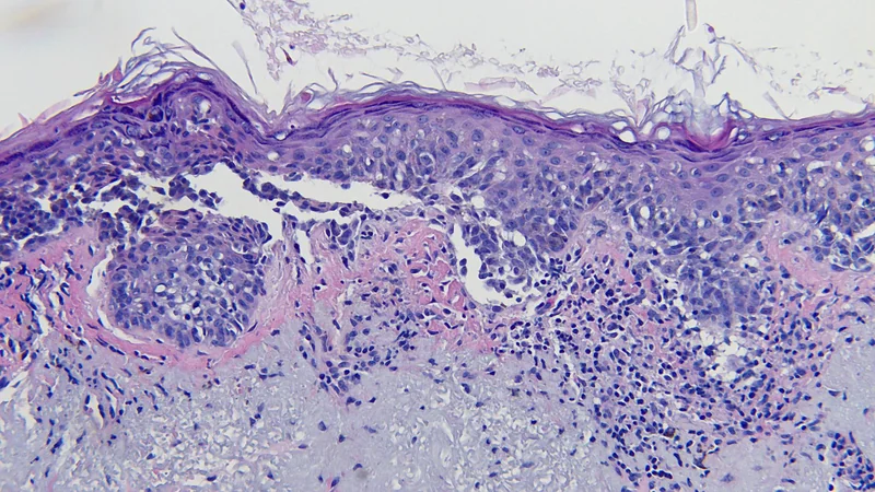 Histology
Definition
Lentigo Maligna (LM) and Lentigo Maligna Melanoma (LMM)
- Definition: Lentigo maligna is a melanoma in situ on chronically sun-damaged skin; only once the basement membrane is breached is it called lentigo maligna melanoma.
- Clinical picture: a slowly enlarging, ill-defined, irregularly brown-black macule on the chronically sun-exposed face of older patients (cheek, temple, nose).
Differential diagnoses
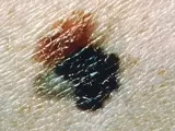 Melanoma
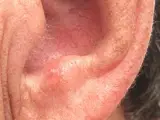 Actinic keratosis
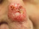 Basal cell carcinoma
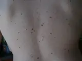 Dysplastic naevus (atypical naevus)
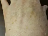 Solar lentigo (age spot)
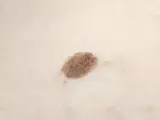 Seborrhoeic keratosis
Image credits for the thumbnails
- Melanoma: National Cancer Institute (unknown photographer), “Melanoma (4)”, Public Domain, via Wikimedia Commons · Public domain · Source
- Actinic keratosis: Future FamDoc, “Actinic keratosis on ear”, CC BY-SA 4.0, via Wikimedia Commons · CC BY-SA 4.0 · Source
- Basal cell carcinoma: Bin im Garten, “Basaliom am Nasenrücken 2”, CC BY-SA 3.0, via Wikimedia Commons · CC BY-SA 3.0 · Source
- Dysplastic naevus (atypical naevus): 0x6adb015 (Wikimedia Commons), “DysplasticNevusSyndrome”, CC BY-SA 3.0, via Wikimedia Commons · CC BY-SA 3.0 · Source
- Solar lentigo (age spot): Beyond My Ken, “Liver spots, light-skinned Caucasian man, age 63”, CC BY-SA 4.0, via Wikimedia Commons · CC BY-SA 4.0 · Source
- Seborrhoeic keratosis: Assafn, “Seborrheic keratosis closup”, CC BY-SA 4.0, via Wikimedia Commons · CC BY-SA 4.0 · Source
Treatment principles
- The treatment of choice is complete excision with complete histological margin control (micrographically controlled surgery), as the extent is often clinically underestimated.
- In patients unfit for surgery or at unfavourable sites, radiotherapy may be considered; other non-surgical procedures are individual case decisions.
- Because of the risk of progression to lentigo maligna melanoma and of recurrence, long-term follow-up and sun protection are required.
Guideline: AWMF guideline register (no. 032-024OL)
Specific treatment regimens with drugs and doses are covered in the DermaFuchs app.
Keep learning in the app
References (selection)
Cross-references
Keep learning
Keep learning with flashcards, exam questions and spot diagnoses in the DermaFuchs app – free, in your browser or as an app.
Related pages: Melanoma · Actinic keratosis · Basal cell carcinoma
Note: This page is intended for medical education and does not replace diagnosis or treatment decisions in individual cases.