Actinic keratosis

Synonyms
solare Keratose, Präkanzerose, AK
Specialty
Dermatology · Skin tumours
Images
Clinical 4 · Histology 3
In the app
9 flashcards
Last updated
09/2026 · Dr. Pascal Bafteh
Contents
  1. Images (7)
  2. Definition
  3. Classification
  4. Aetiopathogenesis
  5. Differential diagnoses
  6. Keep learning in the app
  7. References (selection)
  8. Cross-references

Images (7)

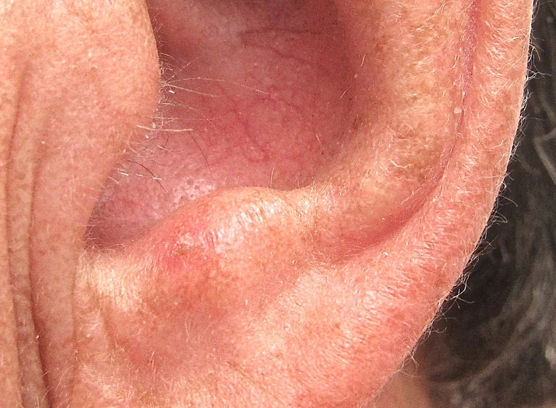
Actinic keratosis – Clinical: rough, erythematous, scaly plaque on the sun-exposed ear helix
rough, erythematous, scaly plaque on the sun-exposed ear helixFuture FamDoc, „Actinic keratosis on ear", CC BY-SA 4.0, via Wikimedia Commons · CC BY-SA 4.0 · Source
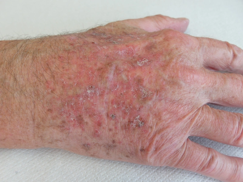
Actinic keratosis – Clinical: thick yellowish-white hyperkeratosis on an erythematous base with field cancerisation. (Photo: Pascal Bafteh)
thick yellowish-white hyperkeratosis on an erythematous base with field cancerisation. (Photo: Pascal Bafteh)Photo: Dr. Pascal Bafteh (own practice, published with patient consent)
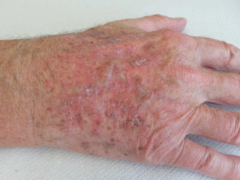
Actinic keratosis – Clinical: multiple rough, scaly reddish lesions on a sun-damaged back of the hand (field cancerization) (Photo: Dr. Thomas Brinkmeier, Wikimedia Commons, CC BY 4.0)
multiple rough, scaly reddish lesions on a sun-damaged back of the hand (field cancerization) (Photo: Dr. Thomas Brinkmeier, Wikimedia Commons, CC BY 4.0)Dr. Thomas Brinkmeier, „Aktinische Keratosen am Handrücken, sog. Feldkanzerisierung, ©WIKIDERM", CC BY 4.0, via Wikimedia Commons · CC BY 4.0 · Source
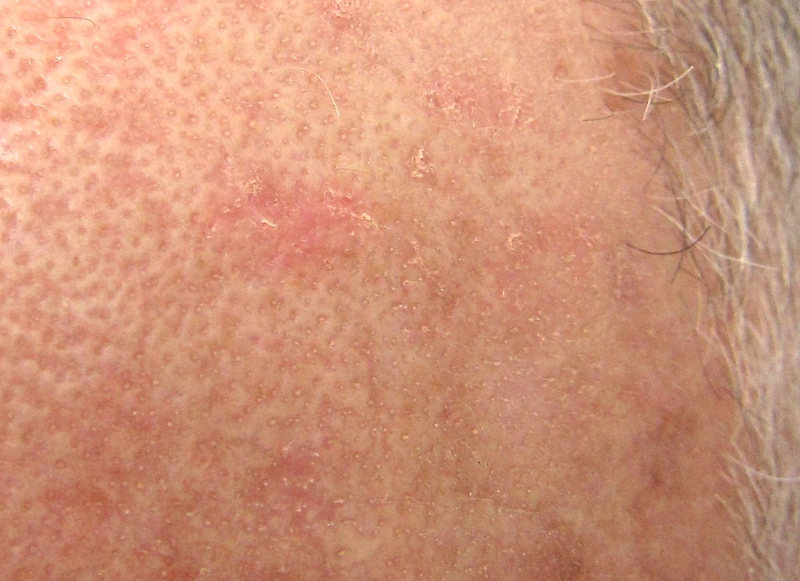
Actinic keratosis – Clinical
Future FamDoc, „Close view of actinic keratosis", CC BY-SA 4.0, via Wikimedia Commons · CC BY-SA 4.0 · Source
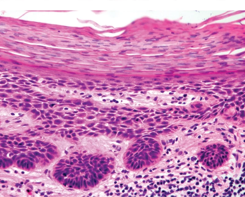
Actinic keratosis – Histology: histology (H&E): a broad band of parakeratosis over crowded atypical keratinocytes with nuclear enlargement and hyperchromasia and loss of orderly maturation, basaloid buds at the …Histology
histology (H&E): a broad band of parakeratosis over crowded atypical keratinocytes with nuclear enlargement and hyperchromasia and loss of orderly maturation, basaloid buds at the …Valerie R. Yanofsky, Stephen E.Mercer, and Robert G. Phelps, „Micrograph of established actinic keratosis", CC BY 4.0, via Wikimedia Commons · CC BY 4.0 · Source
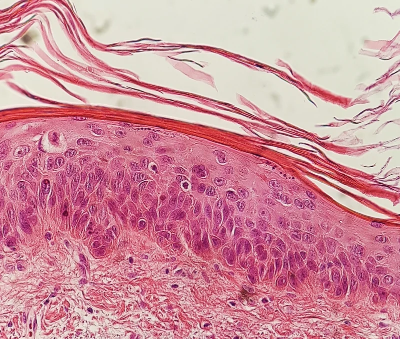
Actinic keratosis – Histology: histology (H&E): atypical keratinocytes from the basal layer extending through about half of the stratum spinosum (moderate atypia)Histology
histology (H&E): atypical keratinocytes from the basal layer extending through about half of the stratum spinosum (moderate atypia)Mikael Häggström, M.D., „Histopathology of actinic keratosis with moderate atypia", CC0, via Wikimedia Commons · CC0 · Source
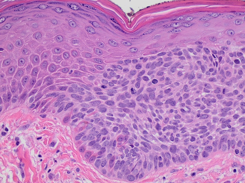
Actinic keratosis – Histology: histology (H&E): normal skin on the left, atypical basal keratinocytes not involving the full epidermal thickness on the rightHistology
histology (H&E): normal skin on the left, atypical basal keratinocytes not involving the full epidermal thickness on the rightMikael Häggström, M.D., „Histopathology of actinic keratosis", CC0, via Wikimedia Commons · CC0 · Source
1 / 7

Definition

Actinic keratosis is a chronic UV-induced dysplasia of the [keratinocytes|fibroblasts|melanocytes|endothelial cells] with potential for progression to a Squamous Cell Carcinoma (SCC) — it is therefore regarded as a carcinoma in situ / early form of SCC and not as a harmless sign of ageing.

Malignancy Risk & Occupational Disease

  • Malignancy risk: Approx. [10|1|5|30] % progression to invasive SCC over [10|2|20|50] years; crucially, even low-grade (Olsen I) lesions can progress to SCC — the clinical grade does not allow any conclusion about the progression risk of the individual lesion.

Classification

Subtypes of Actinic Keratosis

  • [Classical|Pigmented|Bowenoid|Lichenoid] (erythematosquamous) — flat, scaling.
  • Hyperkeratotic — highly keratinized.
  • Atrophic: thin, barely palpable.
  • Lichenoid — inflammatory infiltrated.
  • Pigmented — brownish.
  • Bowenoid — almost complete epidermal involvement.
  • Actinic cheilitis — UV-induced dysplasia of the vermilion border.

Aetiopathogenesis

Risk Factors and Clinical Findings

Predilection sites are the chronically light-exposed "sun terraces": bald scalp, forehead, nose, helix of the ear, cheeks, lower lip, backs of the hands, forearms and lower legs. Clinically they present as erythematous, rough macules and plaques that are characteristically better palpable than visible (sandpaper feel) — palpation of the entire field is therefore the decisive examination step.

Differential diagnoses

Keep learning in the app

In the DermaFuchs app you can learn Actinic keratosis with flashcards, exam questions and spot-the-diagnosis – free, in your browser or as an app.

Test yourself: Spot-diagnosis quiz with real cases →

In the app: 9 flashcards

Open in browser  Download on the App Store

References (selection)

  1. AWMF S3 AK 2023
  2. Long et al. 2023

Cross-references

Note: This page is intended for medical education and does not replace diagnosis or treatment decisions in individual cases.