Amelanotic melanoma
- Synonyms
- amelanotisches malignes Melanom, hypomelanotisches Melanom, pigmentarmes Melanom, Hypomelanotic melanoma
- Specialty
- Dermatology · Skin tumours
- Images
- Clinical 1 · Histology 1
- In the app
- 5 flashcards
- Last updated
- 10/2026 · Dr. Pascal Bafteh
Contents
Images (2)
 Histology
Definition
In amelanotic melanoma the tumour cells produce little or no pigment. The term is often also used for melanomas that are only partly devoid of pigment (hypomelanotic); completely pigment-free melanomas are rare. Any melanoma subtype can be amelanotic.
Occurrence & epidemiology
Depending on the definition, about 2–20% of all melanomas are classed as amelanotic. Risk factors are older age, chronically sun-damaged skin, fair skin, red hair, marked freckling and a tendency to sunburn; amelanotic melanoma also accounts for a considerable proportion of melanoma in children. About 25% of subungual melanomas and over 50% of desmoplastic melanomas are amelanotic.
Clinical features
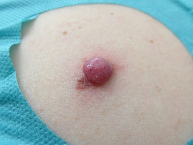
- Skin-coloured, pink or red macules, plaques or nodules, sometimes scaly; at the edge there may be faint light tan or grey pigmentation; borders well or ill defined.
- Nodular forms resemble a skin-coloured naevus or basal cell carcinoma; other mimics include pyogenic granuloma and keratoacanthoma.
- The ABCDE criteria are often not met; asking about red, raised, recent change helps.
- Melanoma metastases may be amelanotic even when the primary tumour was pigmented.
Diagnosis
- History of a new or changing lesion and examination of the entire skin.
- Dermoscopy: the vascular pattern is decisive – polymorphous vessels, most frequently a combination of dotted and linear irregular vessels, milky-red areas; possibly remnants of irregular pigmentation.
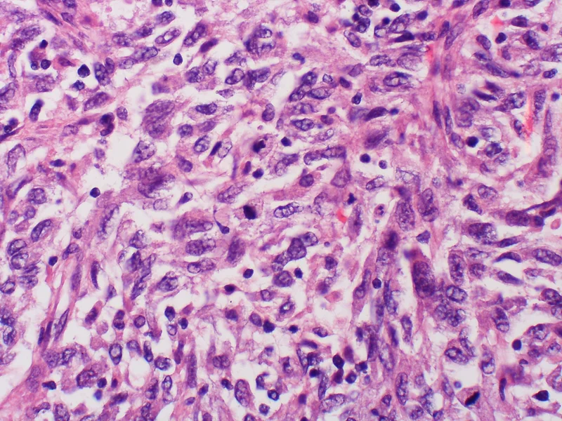
- Histology confirms the diagnosis. The absence of melanin granules (e.g. on Fontana-Masson staining) makes classification harder; immunohistochemistry demonstrates the melanocytic origin. The report includes Breslow thickness, ulceration and mitotic rate, among other features.
Differential diagnoses
Keep learning in the app
References (selection)
Cross-references
Note: This page is intended for medical education and does not replace diagnosis or treatment decisions in individual cases.