Pleomorphic dermal sarcoma
- Synonyms
- PDS
- Specialty
- Dermatology · Skin tumours
- Images
- Clinical 2
- In the app
- 2 flashcards
- Last updated
- 10/2026 · Dr. Pascal Bafteh
Contents
Images (2)
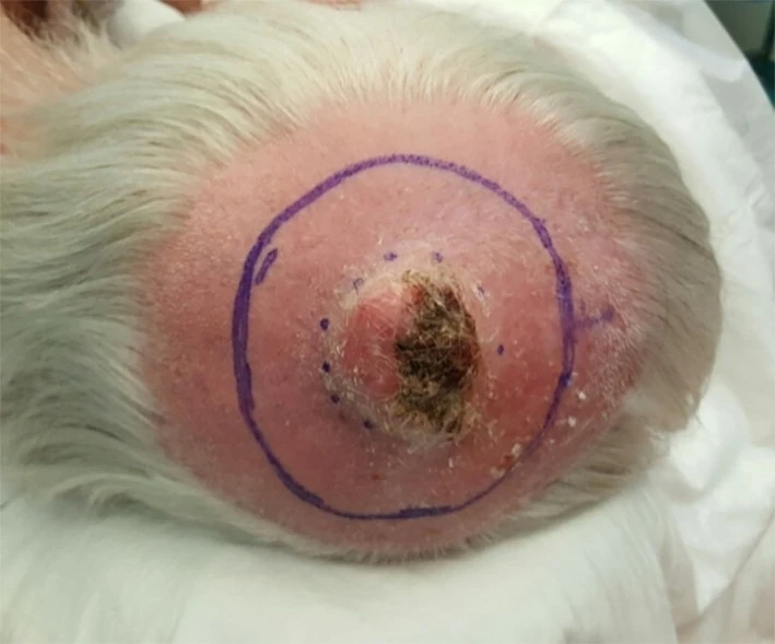
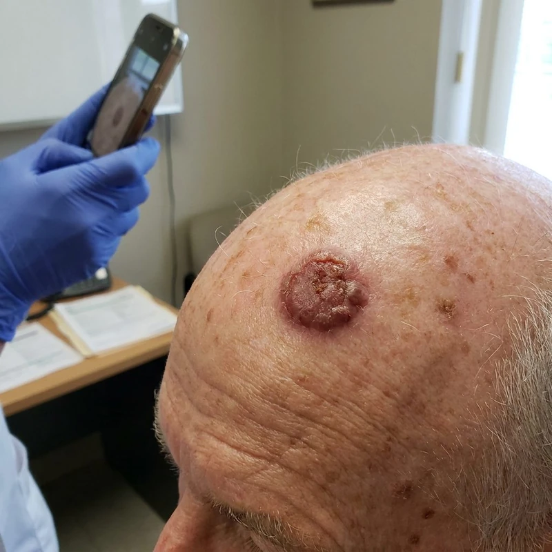 AI illustration
Definition
Pleomorphic Dermal Sarcoma (PDS)
Definition & Differentiation from AFX
- PDS = the "more aggressive / deeper brother" of AFX with at least one aggressive histological feature.
- Definition criteria (one is sufficient): subcutaneous invasion + necrosis + LVI/PNI.
Clinical Presentation
- Clinically like AFX, but poorly circumscribed with infiltrative growth; both fast and slow growth possible.
- Localization: same UV-exposed areas (scalp, cheeks, ears).
Histology
- In addition to AFX: subcutaneous involvement + necrosis + LVI/PNI — all 3 are absent in AFX.
- IHC identical to AFX (CD10+, cytokeratins−, S100/SOX10−, desmin−).
Differential diagnoses
 Dermatofibrosarcoma protuberans
 Melanoma
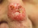 Basal cell carcinoma
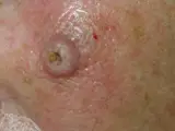 Keratoacanthoma
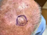 Atypical Fibroxanthoma (AFX)
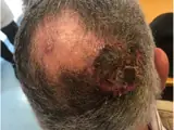 Cutaneous angiosarcoma
Image credits for the thumbnails
- Dermatofibrosarcoma protuberans: Tontonflingueur, “Dermatofibrosarcome facial 01”, CC BY-SA 4.0, via Wikimedia Commons · CC BY-SA 4.0 · Source
- Melanoma: National Cancer Institute (unknown photographer), “Melanoma (4)”, Public Domain, via Wikimedia Commons · Public domain · Source
- Basal cell carcinoma: Bin im Garten, “Basaliom am Nasenrücken 2”, CC BY-SA 3.0, via Wikimedia Commons · CC BY-SA 3.0 · Source
- Keratoacanthoma: Klaus D. Peter, Wiehl, Germany, “Spinaliom”, CC BY 3.0 DE, via Wikimedia Commons · CC BY 3.0 DE · Source
- Atypical Fibroxanthoma (AFX): Apichoto-Mata I, García-Contreras SJ, Garnica B (Cureus 2026), CC BY 4.0, via PubMed Central (PMC12949712) · CC BY 4.0 · Source
- Cutaneous angiosarcoma: Messias H, Martins M, Zagalo C et al. (Cancer Rep (Hoboken) 2022), CC BY 4.0, via PubMed Central (PMC9575495) · CC BY 4.0 · Source
Treatment principles
- The treatment of choice is complete excision with adequate safety margins and deep resection, preferably with micrographic margin control.
- Adjuvant radiotherapy is considered after incomplete resection or in the presence of high-risk features.
- Given the relevant rates of recurrence and metastasis, tumour-board discussion and close follow-up are indicated; there is no established systemic standard therapy for inoperable or metastatic disease, and immune checkpoint inhibitors may be considered in individual cases.
Guideline: AWMF guideline register (no. 032-057)
Specific treatment regimens with drugs and doses are covered in the DermaFuchs app.
Keep learning in the app
Cross-references
Keep learning
Keep learning with flashcards, exam questions and spot diagnoses in the DermaFuchs app – free, in your browser or as an app.
Related pages: Dermatofibrosarcoma protuberans · Melanoma · Basal cell carcinoma
Note: This page is intended for medical education and does not replace diagnosis or treatment decisions in individual cases.