Lipoma
- Synonyms
- Lipome, Fettgewebsgeschwulst, Fettgeschwulst, Angiolipom, Spindelzelllipom, fatty lump, angiolipoma
- Specialty
- Dermatology · Skin tumours
- Images
- Clinical 2 · Histology 1
- Last updated
- 10/2026 · Dr. Pascal Bafteh
Contents
Images (3)
 Histology
Definition
A lipoma is a very common, benign, slow-growing tumour of mature fat cells (adipocytes). It usually lies in the subcutaneous fat, rarely deep to the fascia or within muscle. Malignant transformation is exceedingly rare.
Classification
- Variants: angiolipoma (often painful), spindle cell lipoma (especially on the neck of older people), pleomorphic lipoma, adenolipoma.
- Multiple lipomas (in about 5–10% of patients) are usually part of a syndrome or genetic disorder, e.g. familial multiple lipomatosis, Proteus syndrome, PTEN hamartoma syndrome, Gardner syndrome or MEN 2B; multiple painful lipomas occur in Dercum disease.
Occurrence & epidemiology
Lipomas typically appear between 40 and 60 years of age but can occur at any age. Both sexes are equally affected; they are more common in hyperlipidaemia, type 2 diabetes and obesity. The cause is unknown.
Clinical features
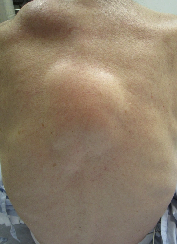
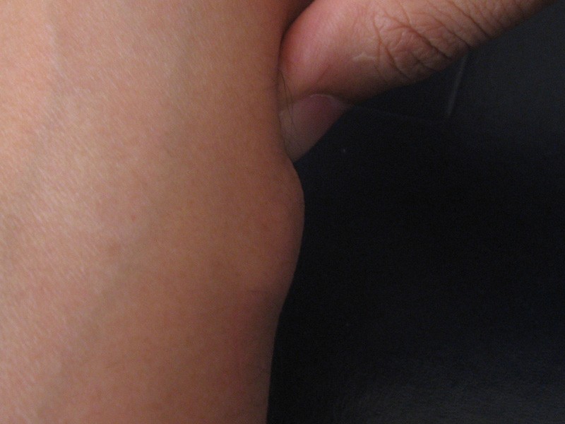
- Usually a solitary, painless, soft to rubbery, often lobulated and freely mobile nodule, 1 to more than 10 cm in diameter.
- The overlying skin is unchanged and not tethered to the nodule.
- Mainly on the trunk, neck and proximal limbs.
- Rarely, large lipomas compress nerves or adjacent structures and cause pain or numbness.
Histology
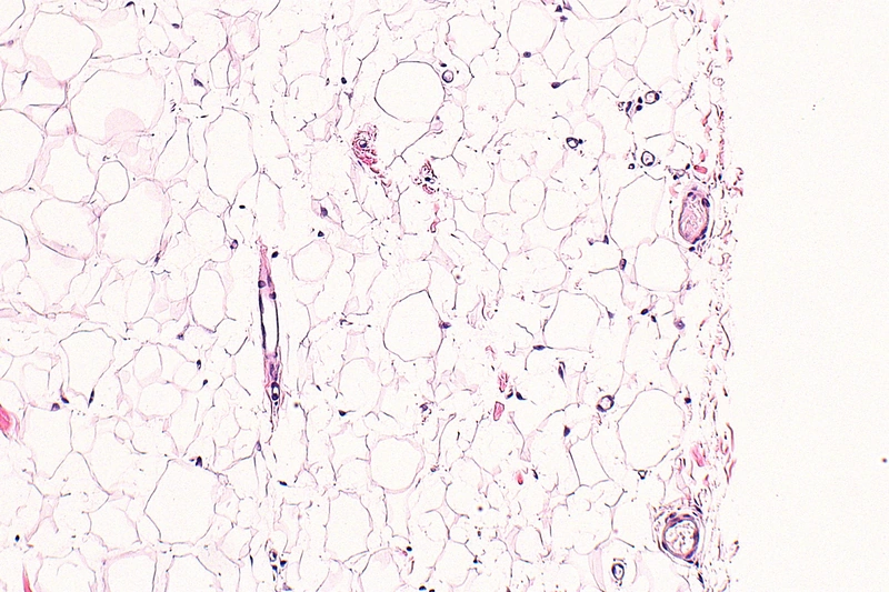
Mature adipose tissue with a few small capillaries in thin fibrous septa, often surrounded by a thin fibrous capsule. Distinguishing it from well-differentiated liposarcoma can be histologically difficult.
Diagnosis
- Usually a clinical diagnosis.
- Imaging and/or biopsy if the findings are uncertain.
- Rapidly growing nodules are examined histologically to exclude liposarcoma.
Differential diagnoses
- Epidermal cyst (atheroma)
- Lipoid proteinosis (Urbach–Wiethe disease)
- Dermatofibroma
- Fibrofolliculoma (Birt-Hogg-Dubé)
- Steatocystoma multiplex
- Abscess
- Haemangioma
Keep learning in the app
References (selection)
Cross-references
Note: This page is intended for medical education and does not replace diagnosis or treatment decisions in individual cases.