Bowen's disease (squamous cell carcinoma in situ)
Exam relevance: in 3 of more than 50 board exam reports · rank 45
- Synonyms
- Bowen-Krankheit, Carcinoma in situ, intraepidermales Plattenepithelkarzinom, Erythroplasie Queyrat, Bowen's disease, squamous cell carcinoma in situ, intraepidermal carcinoma
- Specialty
- Dermatology · Skin tumours
- Images
- Clinical 5 · Histology 3
- In the app
- 10 flashcards
- Last updated
- 10/2026 · Dr. Pascal Bafteh
Contents
Images (8)
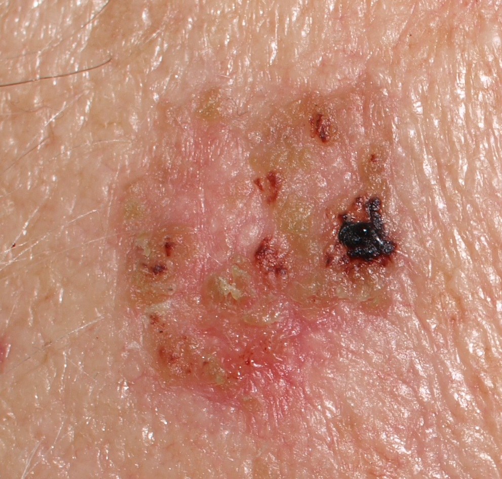
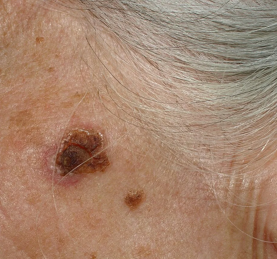
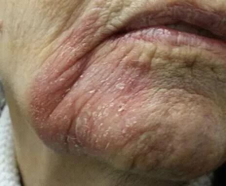
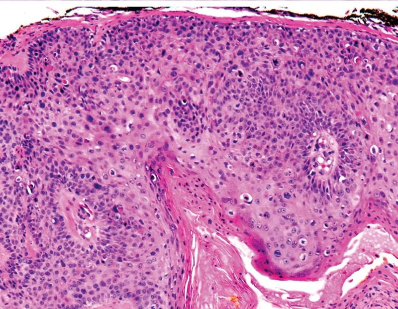 Histology
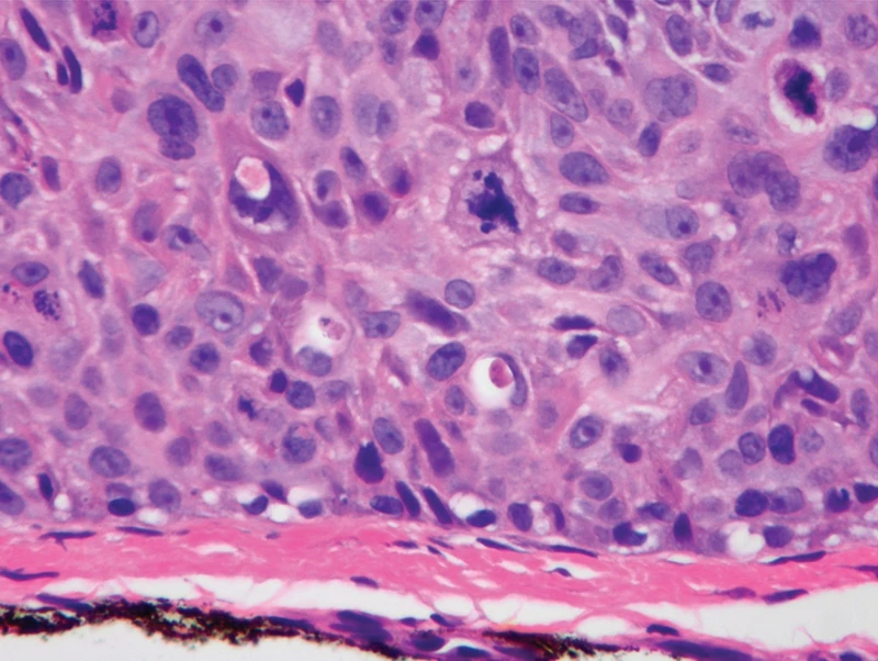 Histology
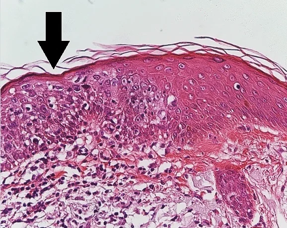 Histology
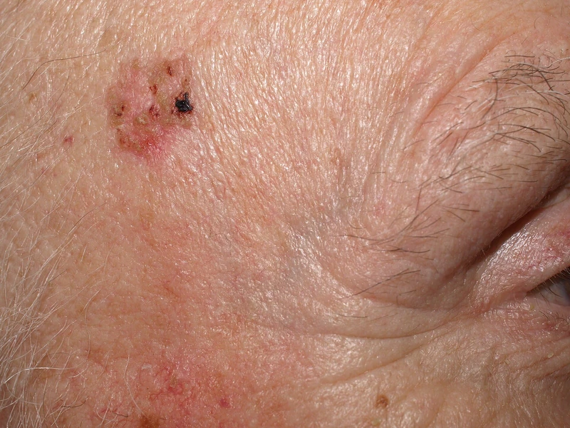
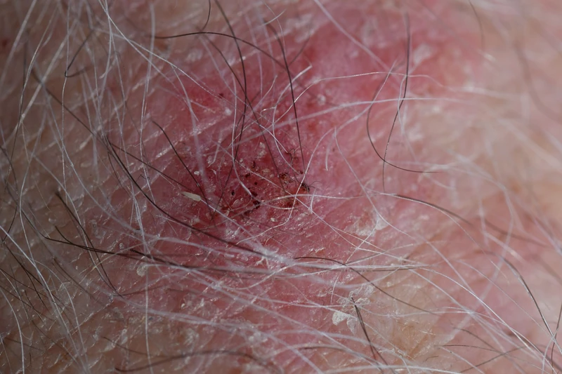
Definition
Bowen's disease is a squamous cell carcinoma in situ of the skin (intraepidermal squamous cell carcinoma). Atypical keratinocytes occupy the full thickness of the epidermis while the basement membrane remains intact. On the mucosa of the glans penis it is called erythroplasia of Queyrat.
Occurrence & epidemiology
Bowen's disease is most often found in sun-damaged skin. Further risk factors are arsenic ingestion, ionising rays, human papillomavirus infection (on fingers and nails) and immunosuppression. Up to half of patients have other keratinocytic skin cancers. Invasive squamous cell carcinoma arises in about 5% of lesions.
Clinical features
- One or more irregular scaly plaques up to several centimetres across, often orange-red, sometimes brown; when multiple, the distribution is asymmetrical (unlike psoriasis).
- Mainly sun-exposed sites (ears, face, hands, lower legs), but possible on any skin; under the nail as a red longitudinal streak (erythronychia) that may later destroy the nail plate.
- Variants: pigmented, pagetoid and clear cell Bowen's disease (defined histologically).
Histology
Atypical keratinocytes across the full thickness of the epidermis with disorderly maturation, loss of the granular layer and overlying parakeratosis; the basement membrane is preserved, and the adnexal ostial epithelium is commonly spared.
Diagnosis
- Dermoscopy: clustered rounded and coiled (glomerular) vessels, white-yellow scale.
- Biopsy for histological confirmation (full-thickness dysplasia of the epidermis), among other things to distinguish psoriasis and invasive squamous cell carcinoma.
Differential diagnoses
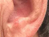 Actinic keratosis
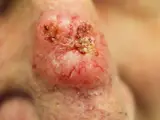 Basal cell carcinoma
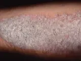 Psoriasis vulgaris (plaque psoriasis)
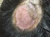 Discoid lupus erythematosus (CDLE)
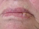 Cutaneous squamous cell carcinoma
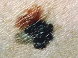 Melanoma
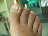 Onychomycosis (nail fungus)
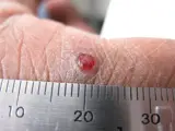 Pyogenic granuloma
Image credits for the thumbnails
- Actinic keratosis: Future FamDoc, “Actinic keratosis on ear”, CC BY-SA 4.0, via Wikimedia Commons · CC BY-SA 4.0 · Source
- Basal cell carcinoma: Bin im Garten, “Basaliom am Nasenrücken 2”, CC BY-SA 3.0, via Wikimedia Commons · CC BY-SA 3.0 · Source
- Psoriasis vulgaris (plaque psoriasis): MediaJet, “An Arm Covered With Plaque Type Psoriasis”, CC BY-SA 3.0, via Wikimedia Commons · CC BY-SA 3.0 · Source
- Discoid lupus erythematosus (CDLE): Mohammad2018, “Discoid lupus etythermayouses new image”, CC BY-SA 4.0, via Wikimedia Commons · CC BY-SA 4.0 · Source
- Cutaneous squamous cell carcinoma: Klaus D. Peter, Wiehl, Germany, “Spinaliom3”, CC BY 3.0 DE, via Wikimedia Commons · CC BY 3.0 DE · Source
- Melanoma: National Cancer Institute (unknown photographer), “Melanoma (4)”, Public Domain, via Wikimedia Commons · Public domain · Source
- Onychomycosis (nail fungus): Bobjgalindo, “Onychomycosis”, CC BY-SA 4.0, via Wikimedia Commons · CC BY-SA 4.0 · Source
- Pyogenic granuloma: kilbad, “Pyogenic granuloma on a finger-1”, CC BY 3.0, via Wikimedia Commons · CC BY 3.0 · Source
Treatment principles
- The treatment of choice is excision with histological examination.
- For multiple or large lesions or at sites with poor wound healing, such as the lower leg, photodynamic therapy, cryotherapy, curettage, topical immunomodulators or cytostatics and radiotherapy may be considered.
- Consistent sun protection and follow-up are important, as progression to invasive squamous cell carcinoma is possible.
Specific treatment regimens with drugs and doses are covered in the DermaFuchs app.
Keep learning in the app
References (selection)
Cross-references
Mentioned in
More skin tumours
Keep learning
Keep learning with flashcards, exam questions and spot diagnoses in the DermaFuchs app – free, in your browser or as an app.
Related pages: Actinic keratosis · Basal cell carcinoma · Psoriasis vulgaris (plaque psoriasis)
Note: This page is intended for medical education and does not replace diagnosis or treatment decisions in individual cases.