Melanocytic naevus (mole)
- Synonyms
- Muttermal, Leberfleck, Nävus, Pigmentnävus, Nävuszellnävus, Junktionsnävus, dermaler Nävus, mole, melanocytic nevus, melanocytic naevus, birthmark
- Specialty
- Dermatology · Skin tumours
- Images
- Clinical 3 · Dermoscopy 5 · Histology 3
- In the app
- 2 flashcards
- Last updated
- 10/2026 · Dr. Pascal Bafteh
Contents
Images (11)
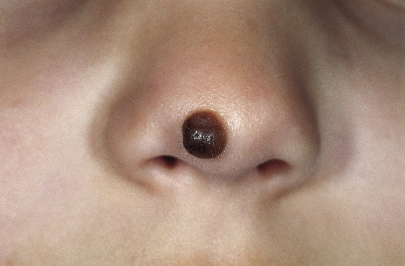
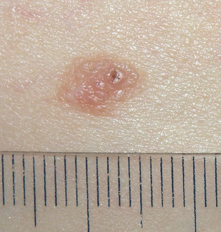
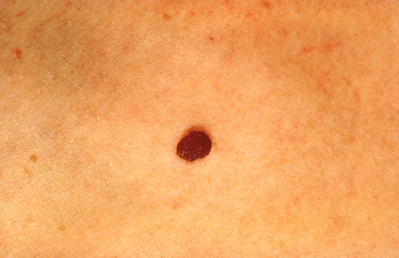
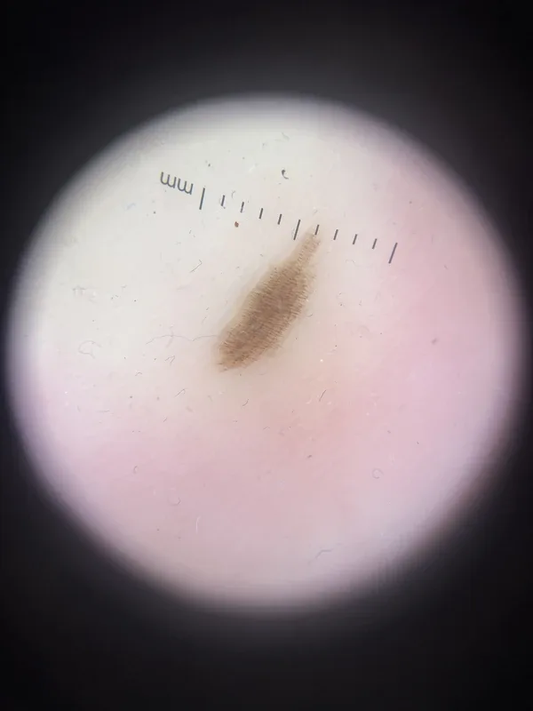 Dermoscopy
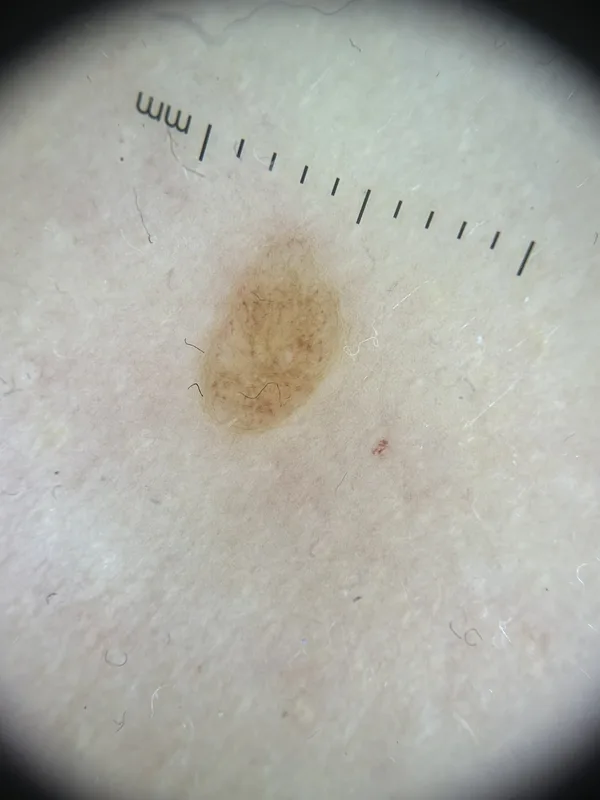 Dermoscopy
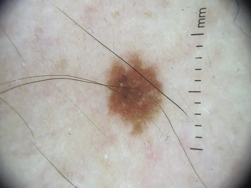 Dermoscopy
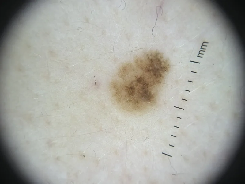 Dermoscopy
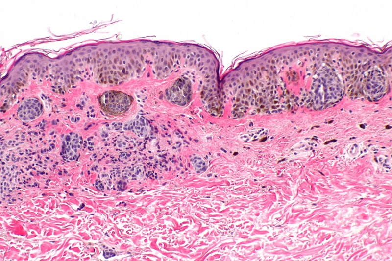 Histology
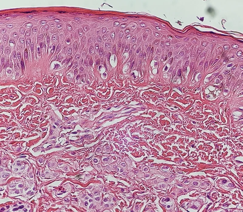 Histology
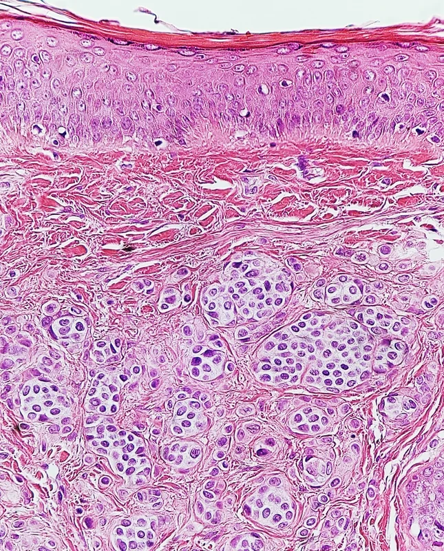 Histology
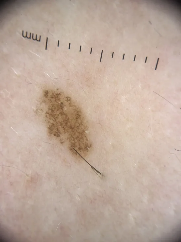 Dermoscopy
Definition
Melanocytic naevi ("moles") are benign, circumscribed collections of naevus cells derived from melanocytes. They may be congenital or acquired during life.
Classification
- By position of the naevus cell nests: junctional naevus (at the dermo-epidermal junction, flat, brown), compound naevus (junctional and dermal, slightly raised), intradermal naevus (purely dermal, skin-coloured to light brown, often soft and papillomatous).
- Congenital naevi: present at birth or appearing shortly afterwards; melanoma risk depends mainly on size – very low for small and medium congenital naevi (well under 1%), increased for giant naevi (20 cm or more).
- Special forms: Spitz naevus, blue naevus, halo naevus, dysplastic (atypical) naevus, cockade naevus.
Occurrence & epidemiology
Naevi appearing in childhood (about age 2–10) tend to be the most prominent and persist for life; most fair-skinned adults have 20–50 naevi. The number depends on genetic factors, sun exposure and immune status; fair-skinned people have more naevi. A high naevus count (especially over 100) and atypical naevi are important risk markers for melanoma.
Clinical features
- Round to oval, symmetrical, sharply demarcated, evenly pigmented macules or papules.
- Warning signs of melanoma: ABCD features (asymmetry, border irregularity, colour variation, diameter over 6 mm), change in size, shape, structure or colour, a new naevus in adult life (over 40 years) and bleeding, crusting or itch.
- "Ugly duckling sign": a naevus that looks clearly different from the patient's other naevi.
Diagnosis
- Total body examination and dermoscopy (regular pigment network, globules or homogeneous pattern; symmetrical distribution of colours and structures).
- Digital follow-up imaging for many or atypical naevi; histological examination if melanoma is suspected.
Differential diagnoses
 Melanoma
 Dysplastic naevus (atypical naevus)
 Seborrhoeic keratosis
 Spitz naevus (spindle and epithelioid cell naevus)
 Blue naevus
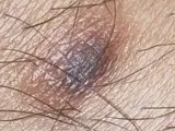 Dermatofibroma
Image credits for the thumbnails
- Melanoma: National Cancer Institute (unknown photographer), “Melanoma (4)”, Public Domain, via Wikimedia Commons · Public domain · Source
- Dysplastic naevus (atypical naevus): 0x6adb015 (Wikimedia Commons), “DysplasticNevusSyndrome”, CC BY-SA 3.0, via Wikimedia Commons · CC BY-SA 3.0 · Source
- Seborrhoeic keratosis: Assafn, “Seborrheic keratosis closup”, CC BY-SA 4.0, via Wikimedia Commons · CC BY-SA 4.0 · Source
- Spitz naevus (spindle and epithelioid cell naevus): The Armed Forces Institute of Pathology (AFIP), Public domain, via Wikimedia Commons · Public domain · Source
- Blue naevus: Dannii Brighton, “Blue Nevus”, CC BY-SA 3.0, via Wikimedia Commons · CC BY-SA 3.0 · Source
- Dermatofibroma: Jmarchn, “Pigmented and tenderness dermatofibroma”, CC BY-SA 3.0, via Wikimedia Commons · CC BY-SA 3.0 · Source
Treatment principles
- Common melanocytic naevi do not require treatment.
- Clinically or dermoscopically suspicious naevi are completely excised and examined histologically; naevi removed for cosmetic reasons should also undergo histology.
- Ablative procedures without histology, such as laser, are not appropriate for melanocytic naevi; sun protection and, in patients with many naevi, regular skin cancer screening are advisable.
Specific treatment regimens with drugs and doses are covered in the DermaFuchs app.
Keep learning in the app
References (selection)
Cross-references
Mentioned in
Keep learning
Keep learning with flashcards, exam questions and spot diagnoses in the DermaFuchs app – free, in your browser or as an app.
Related pages: Melanoma · Dysplastic naevus (atypical naevus) · Seborrhoeic keratosis
Note: This page is intended for medical education and does not replace diagnosis or treatment decisions in individual cases.