Basal cell carcinoma
General
Basal cell carcinoma Origin: follicular/perifollicular pluripotent stem cells, Mutation of PTCH1/SMO in the Hedgehog signaling pathway or p53.
- Incidence: approx. 1:1000, semi-malignant, metastasis approx. 0.0025%.
- Special form: Pinkus tumor (Fibroepithelioma).
Risk Factors
General Risk Factors
- RF: UV exposure, arsenic, fair skin type, noxious agents, radiation.
- Further RF: Albinism, Xeroderma pigmentosum.
- Medications: AZA, Ciclosporin, MTX, TNF-α, Calcineurin inhibitors.
Basal Cell Carcinoma Syndrome (Gorlin-Goltz)
- Diagnostic criteria: multiple BCCs <30 years, palmar pits, jaw cysts, calcified falx, BCC in first-degree relatives, flat nasal root.
- Important: no radiation, rather PDT.
Clinical Presentation
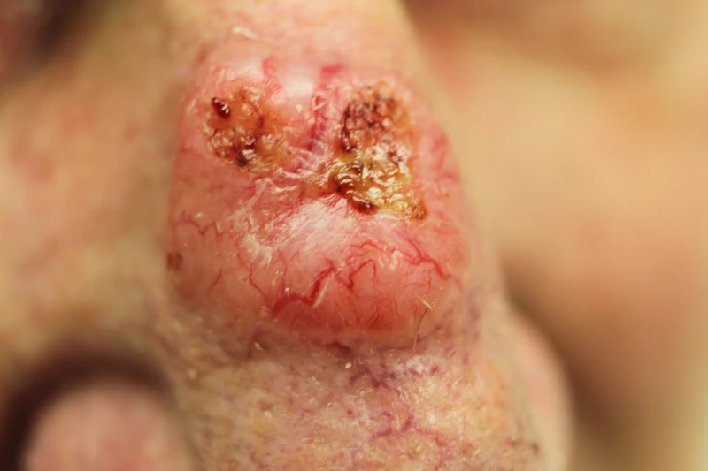
- Lesions: Skin-colored or red or reddish-brown papules, plaques, nodules, or ulcers.
- Typical: pearled border and telangiectasias.
- Further signs: Surface sheen, bizarre tumor vessels.
Dermoscopy of BCC
- Entry criterion: A BCC shows no pigment network — only once a network is absent are the BCC criteria applied at all.
- Vascular criterion: arborizing (tree-like branching) telangiectasias — bold, sharply focused vessels branching dichotomously on the tumour surface. Superficial BCC instead shows short, fine telangiectasias.
- Pigment criteria (especially pigmented BCC): blue-grey ovoid nests (large, sharply demarcated pigment aggregates), multiple blue-grey globules, maple-leaf-like areas at the periphery, spoke-wheel areas and concentric structures.
- Further signs: ulceration or multiple small erosions, and shiny white blotches and strands (visible only under polarized light).
- Note: spoke-wheel areas and arborizing vessels are highly specific; the absent pigment network separates pigmented BCC from melanoma.
Clinical Subtypes
- Nodular BCC: most common type; pearly papule with telangiectasias, usually face.
- Superficial BCC: erythematous, sharply demarcated plaque, usually trunk.
- Sclerodermiform (morpheaform) BCC: scar-like, infiltrative, ill-defined margin, high recurrence risk.
- Pigmented BCC: darkly pigmented; DD: melanoma.
- Infiltrative BCC: aggressive growth, ill-defined border.
- Micronodular BCC: small tumor nests, aggressive.
- Fibroepithelioma of Pinkus: lumbosacral, skin-colored papule, benign BCC variant.
- Basosquamous carcinoma: mixed BCC + SCC features, higher metastatic potential.
Differential diagnoses
- Keratoacanthoma
- Dermatofibroma
- Melanoma
- Trichoepithelioma
- Actinic keratosis
- Seborrhoeic keratosis
- Cutaneous squamous cell carcinoma
- Sebaceous hyperplasia
Practise Basal cell carcinoma in the app
Flashcards with spaced repetition, exam questions and spot-the-diagnosis on this topic – in DermaFuchs, free of charge.
In the DermaFuchs app: 17 flashcards · 27 clinical images
Open in browser Download on the App StoreSources (selection)
More skin tumours
- Melanoma
- Lentigo maligna
- Cutaneous squamous cell carcinoma
- Actinic keratosis
- Actinic cheilitis
- Merkel cell carcinoma
- Microcystic adnexal carcinoma
- Dermatofibroma
Note: This page is intended for medical education and does not replace diagnosis or treatment decisions in individual cases. Treatment and follow-up content is available in the app.