Xeroderma pigmentosum (XP)
- Synonyms
- xeroderma pigmentosum, XP, children of the moon
- Specialty
- Dermatology · Hereditary skin diseases
- Images
- Clinical 2
- In the app
- 5 flashcards
- Last updated
- 10/2026 · Dr. Pascal Bafteh
Contents
Images (2)
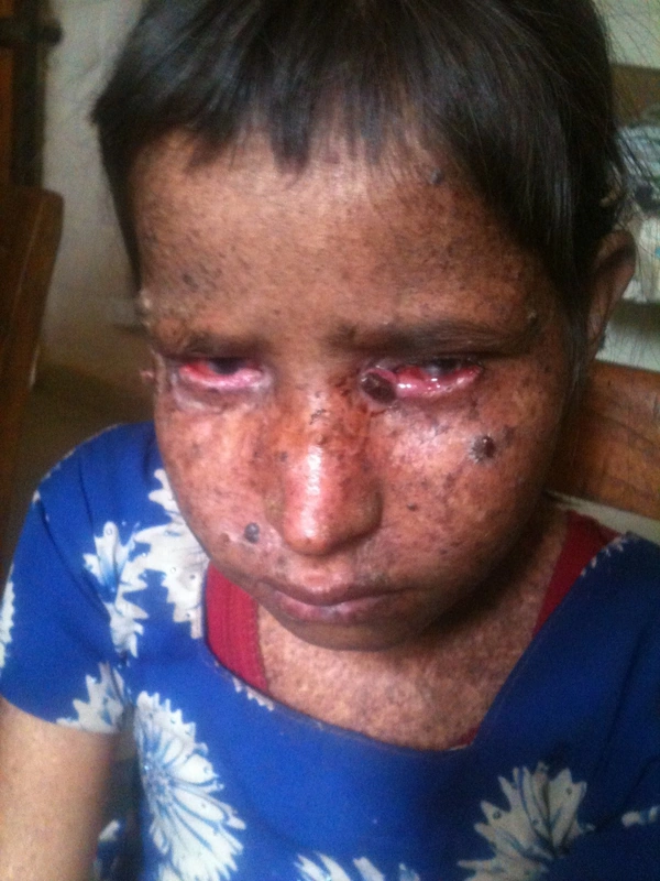
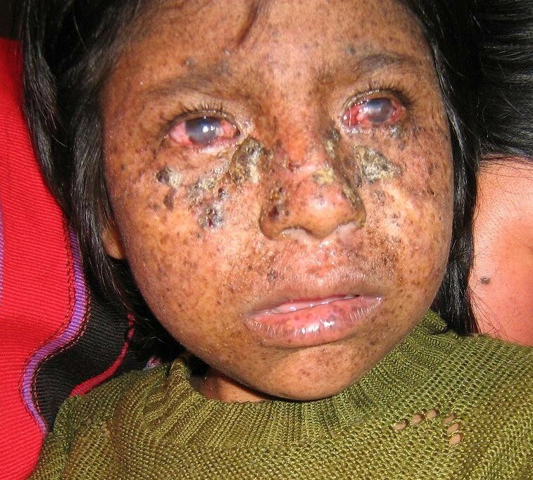
Definition
Xeroderma pigmentosum is a rare autosomal recessive disorder with a defective DNA repair system (NER) that normally cuts out damaged nucleotides. UV-induced DNA damage is not repaired, so that sun damage and skin tumours (basal cell carcinoma, squamous cell carcinoma, melanoma; as early as 4–5 years, mean 8 years) develop already in childhood. At least seven complementation groups (XPA to XPG) are known.
- Genetics: NER defect (pyrimidine dimers), AR. Complementation groups XPA–XPG + XPV (POLη defect).
- Clinical features: Poikiloderma from early childhood, early actinic keratoses, squamous cell and basal cell carcinomas + melanomas (>1000-fold increased!), conjunctivitis + ocular malignancies, neurological deficits (DeSanctis-Cacchione syndrome).
Diagnosis
- Clinical, with striking photosensitivity (severe sunburn after brief exposure) and early poikiloderma with freckling.
- Confirmation by measuring DNA repair capacity in skin or blood samples.
- Eye involvement (photophobia, conjunctivitis, tumours) in nearly 80%, neurological involvement (e.g. spasticity, poor coordination, deafness) in about 20%.
Differential diagnoses
 Actinic keratosis
 Solar lentigo (age spot)
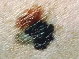 Melanoma
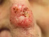 Basal cell carcinoma
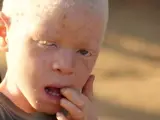 Oculocutaneous albinism
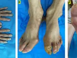 Werner syndrome (adult progeria)
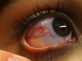 Ataxia-telangiectasia (Louis-Bar syndrome)
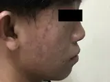 Bloom syndrome
Image credits for the thumbnails
- Actinic keratosis: Future FamDoc, “Actinic keratosis on ear”, CC BY-SA 4.0, via Wikimedia Commons · CC BY-SA 4.0 · Source
- Solar lentigo (age spot): Beyond My Ken, “Liver spots, light-skinned Caucasian man, age 63”, CC BY-SA 4.0, via Wikimedia Commons · CC BY-SA 4.0 · Source
- Melanoma: National Cancer Institute (unknown photographer), “Melanoma (4)”, Public Domain, via Wikimedia Commons · Public domain · Source
- Basal cell carcinoma: Bin im Garten, “Basaliom am Nasenrücken 2”, CC BY-SA 3.0, via Wikimedia Commons · CC BY-SA 3.0 · Source
- Oculocutaneous albinism: Janjacob, “Albino boy by JJ Hoefnagel”, CC BY-SA 3.0, via Wikimedia Commons · CC BY-SA 3.0 · Source
- Werner syndrome (adult progeria): Peng H, Wang J, Liu Y et al. (Front Endocrinol 2022), CC BY 4.0, via PubMed Central (PMC9334726) – panels D–F (cropped) · CC BY 4.0 · Source
- Ataxia-telangiectasia (Louis-Bar syndrome): Thomas O. Crawford, “Photo Ocular telangiectasia in a person with A-T”, CC BY-SA 3.0, via Wikimedia Commons · CC BY-SA 3.0 · Source
- Bloom syndrome: Deng M, Yu M, Jiajue R, Feng K, Xiao X (Front Endocrinol 2021), CC BY 4.0, via PubMed Central (PMC8220076) – figure 1 · CC BY 4.0 · Source
Treatment principles
- There is no causal treatment; lifelong, strict protection from UV radiation through protective clothing, UV-filtering films, sunscreens and avoidance of daylight is essential.
- Close dermatological and ophthalmological follow-up allows early treatment of precancerous lesions and skin tumours, mainly by excision.
- Care is multidisciplinary, including neurological follow-up and genetic counselling.
Specific treatment regimens with drugs and doses are covered in the DermaFuchs app.
Keep learning in the app
References (selection)
Cross-references
Mentioned in
Keep learning
Keep learning with flashcards, exam questions and spot diagnoses in the DermaFuchs app – free, in your browser or as an app.
Related pages: Actinic keratosis · Solar lentigo (age spot) · Melanoma
Note: This page is intended for medical education and does not replace diagnosis or treatment decisions in individual cases.