Seborrhoeic keratosis
Exam relevance: in 1 of more than 50 board exam reports · rank 93
- Synonyms
- Alterswarze, Verruca seborrhoica, Basalzellpapillom, senile Warze, Stuccokeratose, seborrheic keratosis, senile wart, barnacle
- Specialty
- Dermatology · Skin tumours
- Images
- Clinical 2 · Dermoscopy 1 · Histology 4
- In the app
- 3 flashcards
- Last updated
- 10/2026 · Dr. Pascal Bafteh
Contents
Images (7)
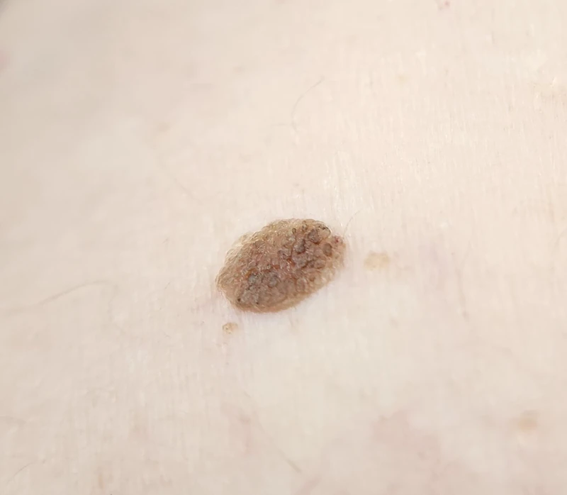
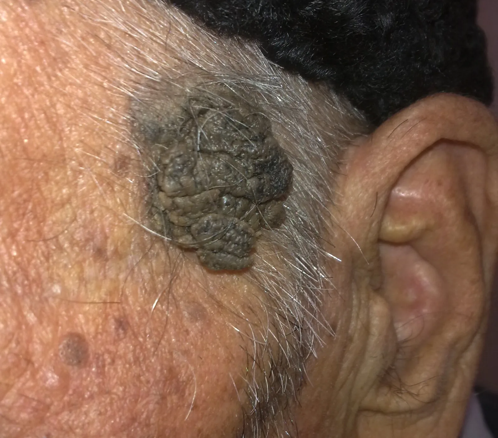
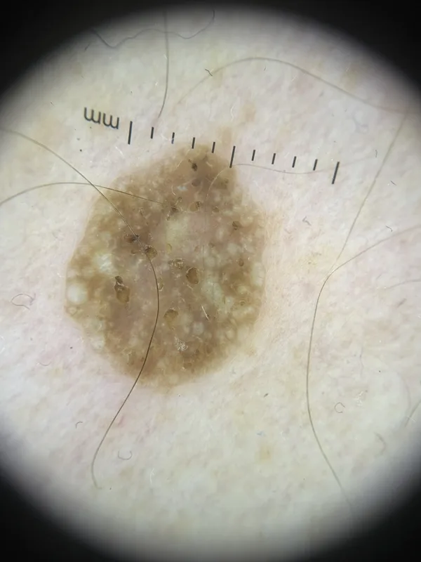 Dermoscopy
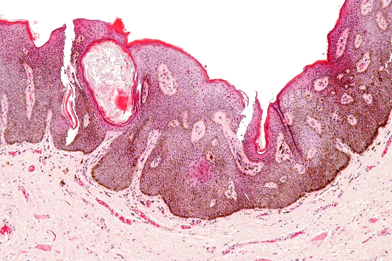 Histology
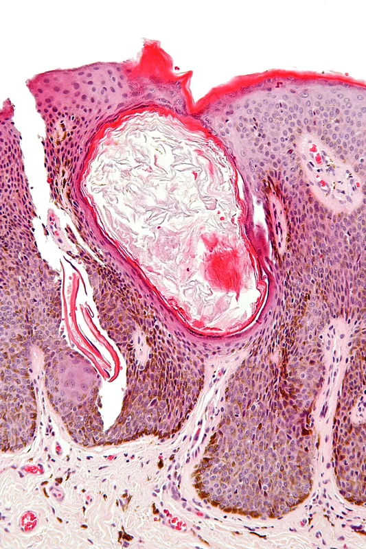 Histology
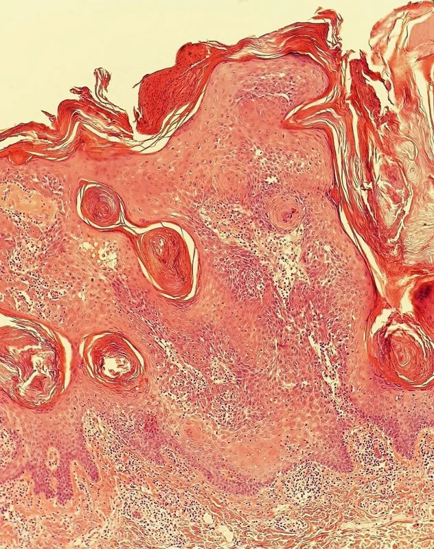 Histology
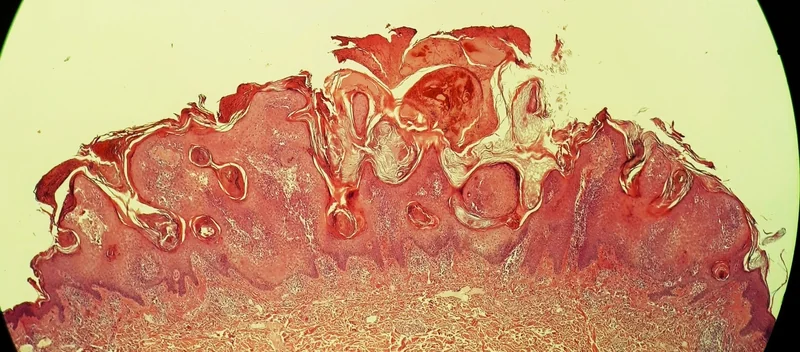 Histology
Definition
Seborrhoeic keratosis (seborrhoeic wart, "senile wart") is a very common benign epidermal tumour and a typical sign of skin ageing. It arises from a proliferation of keratinocytes and is unrelated to wart viruses or sebaceous glands.
Occurrence & epidemiology
Seborrhoeic keratoses typically begin to appear in the 30s or 40s and are uncommon under the age of 20; over 90% of people over 60 have at least one. An inherited tendency to very numerous keratoses occurs. Somatic mutations, for example in FGFR3 and PIK3CA, are found in the lesions. They can arise anywhere except the palms, soles and mucous membranes.
Clinical features
- Sharply demarcated papules and plaques that look "stuck on", skin-coloured, yellow, grey, light to dark brown or black, with a smooth, waxy or warty surface; size from a few millimetres to several centimetres.
- Variants: stucco keratoses (small whitish-grey keratoses on the lower legs and feet), dermatosis papulosa nigra in dark skin, irritated seborrhoeic keratosis with inflammation and crusting.
- Very rarely, the eruptive appearance of numerous seborrhoeic keratoses (sign of Leser-Trélat) points to an internal malignancy, most often gastric adenocarcinoma; eruptive keratoses can also follow sunburn or dermatitis or be drug-induced.
Histology
Well-demarcated proliferation of epidermal keratinocytes with papillomatosis, acanthosis, hyperkeratosis (loose lamellar stratum corneum) and horn cysts; histological patterns include acanthotic, hyperkeratotic/papillomatous, adenoid/reticulated, clonal, irritated and melanoacanthoma.
Diagnosis
- Usually clinical and dermoscopic: white milia-like clods, multiple orange to brown clods (keratin in surface crevices), curved thick ridges and furrows ("brain-like pattern"), sharp border.
- Histology when the distinction is uncertain, especially from melanoma, basal cell or squamous cell carcinoma (collision tumours are possible), or in inflamed lesions.
Differential diagnoses
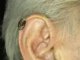
 Actinic keratosis
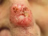 Basal cell carcinoma
 Cutaneous horn (cornu cutaneum)
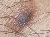 Dermatofibroma
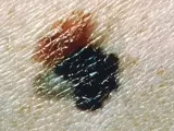 Melanoma
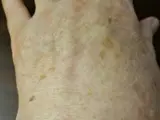 Solar lentigo (age spot)
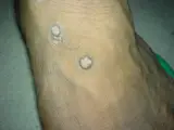 Verruca vulgaris
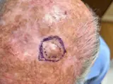 Atypical fibroxanthoma
Image credits for the thumbnails
- Actinic keratosis: Future FamDoc, “Actinic keratosis on ear”, CC BY-SA 4.0, via Wikimedia Commons · CC BY-SA 4.0 · Source
- Basal cell carcinoma: Bin im Garten, “Basaliom am Nasenrücken 2”, CC BY-SA 3.0, via Wikimedia Commons · CC BY-SA 3.0 · Source
- Cutaneous horn (cornu cutaneum): Klaus D. Peter, Wiehl, Germany, “Cornu cutaneum2”, CC BY 3.0 DE, via Wikimedia Commons · CC BY 3.0 DE · Source
- Dermatofibroma: Jmarchn, “Pigmented and tenderness dermatofibroma”, CC BY-SA 3.0, via Wikimedia Commons · CC BY-SA 3.0 · Source
- Melanoma: National Cancer Institute (unknown photographer), “Melanoma (4)”, Public Domain, via Wikimedia Commons · Public domain · Source
- Solar lentigo (age spot): Beyond My Ken, “Liver spots, light-skinned Caucasian man, age 63”, CC BY-SA 4.0, via Wikimedia Commons · CC BY-SA 4.0 · Source
- Verruca vulgaris: Abbassyma (English Wikipedia), “Verruca vulgaris”, Public Domain, via Wikimedia Commons · Public domain · Source
- Atypical fibroxanthoma: Apichoto-Mata I, García-Contreras SJ, Garnica B (Cureus 2026), CC BY 4.0, via PubMed Central (PMC12949712) · CC BY 4.0 · Source
Treatment principles
- Seborrhoeic keratoses are benign and do not require treatment.
- If irritated, itchy or cosmetically troubling, they can be removed by curettage, shave excision, cryotherapy or ablative laser.
- Where there is diagnostic uncertainty, the material should be examined histologically; the sudden eruption of numerous lesions (sign of Leser-Trélat) may warrant a search for malignancy.
Specific treatment regimens with drugs and doses are covered in the DermaFuchs app.
Keep learning in the app
References (selection)
Cross-references
Mentioned in
Keep learning
Keep learning with flashcards, exam questions and spot diagnoses in the DermaFuchs app – free, in your browser or as an app.
Related pages: Actinic keratosis · Basal cell carcinoma · Cutaneous horn (cornu cutaneum)
Note: This page is intended for medical education and does not replace diagnosis or treatment decisions in individual cases.