Dermatofibroma
Definition & Classification
- Dermatofibroma (synonyms: benign fibrous histiocytoma, in everyday practice simply "histiocytoma") = benign fibrohistiocytic proliferation of the dermis.
- Pathogenesis: predominantly interpreted as a reactive fibroblast proliferation after minor trauma (insect bite, thorn injury, shaving); a subset of lesions is nevertheless clonal.
- Epidemiology: peak incidence 20–50 years of age, women more often; one of the most common benign skin tumours overall.
- Dignity: benign, no risk of malignant transformation, no metastasis — its whole importance lies in the distinction from malignant spindle cell tumours.
Clinical Features
- Site: preferentially the lower extremity, especially the lower leg — about two thirds of lesions; women are affected considerably more often.
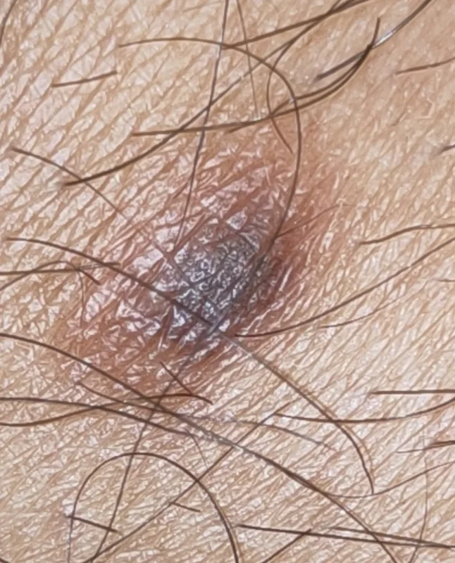
- Morphology: usually a solitary papule or flat nodule of 0.5–1 cm, firm like a lentil, skin-coloured to reddish-brown, often with a peripheral pigmented rim.
- Palpation: the lesion feels firm and button-like and sits within the dermis — it is tethered to the epidermis but mobile over the subcutis.
- Dimple sign (Fitzpatrick sign): on lateral compression between thumb and index finger the lesion dimples inward — a consequence of its anchoring in the fibrosed corium.
- Symptoms: usually asymptomatic, occasionally itching or tenderness; shaving injuries on the lower leg are a frequent reason for presentation.
Dermoscopy
- Classic basic pattern (the most frequent of the described patterns): central whitish scar-like patch with a delicate peripheral pigment network.
- The central white zone corresponds histologically to dermal fibrosis with thickened collagen bundles and is the decisive feature separating it from a melanocytic nevus.
- The peripheral network is fine, light brown and fades out gradually — in the prospective series of 412 lesions by Zaballos the most constant single feature.
- Variants (Zaballos described ten dermoscopic patterns): total or partial pigmentation, vessel-dominated forms with dotted vessels within the white area; atypical patterns do occur and justify excision in case of doubt.
Histology & Immunohistochemistry
- Architecture: an ill-defined dermal proliferation of spindle cells in a storiform-fascicular arrangement, unencapsulated, without tentacular permeation of the subcutaneous fat.
- Collagen trapping: at the periphery the spindle cells surround thick, eosinophilic collagen bundles — a marginal finding typical of dermatofibroma.
- Overlying epidermis: acanthosis with basal hyperpigmentation; follicular and basaloid induction phenomena are also seen.
- Immunohistochemistry: dermatofibroma is factor XIIIa positive, CD34 negative.
- Counter-check dermatofibrosarcoma: dermatofibrosarcoma protuberans (DFSP) shows the mirror image, CD34 positive, factor XIIIa negative, and carries the COL1A1-PDGFB fusion.
Differential diagnoses
- Lipoma
- Epidermal cyst (atheroma)
- Basal cell carcinoma
- Melanoma
- Dermatofibrosarcoma protuberans
Practise Dermatofibroma in the app
Flashcards with spaced repetition, exam questions and spot-the-diagnosis on this topic – in DermaFuchs, free of charge.
In the DermaFuchs app: 3 flashcards · 6 clinical images
Open in browser Download on the App StoreSources (selection)
- Wan L, Park A, Almatroud L, Khachemoune A. Dermatofibroma: Reappraisal and Updated Review. Clin Cosmet Investig Dermatol. 2025;18:1873-1887.
- Luzar B, Calonje E. Cutaneous fibrohistiocytic tumours - an update. Histopathology. 2010;56(1):148-165.
- Zaballos P, Puig S, Llambrich A, Malvehy J. Dermoscopy of dermatofibromas: a prospective morphological study of 412 cases. Arch Dermatol. 2008;144(1):75-83.
- Hardy CSC, Razavi A, Nunez N, et al. Immunohistochemical Profiles of Dermatofibroma and Dermatofibrosarcoma Protuberans: A Scoping Review. Appl Immunohistochem Mol Morphol. 2026;34(1):5-14.
More skin tumours
- Basal cell carcinoma
- Melanoma
- Lentigo maligna
- Cutaneous squamous cell carcinoma
- Actinic keratosis
- Actinic cheilitis
- Merkel cell carcinoma
- Microcystic adnexal carcinoma
Note: This page is intended for medical education and does not replace diagnosis or treatment decisions in individual cases. Treatment and follow-up content is available in the app.