Cutaneous horn (cornu cutaneum)
- Synonyms
- Hauthorn, Keratinhorn, Cornu cutaneum, cutaneous horn, keratin horn
- Specialty
- Dermatology · Skin tumours
- Images
- Clinical 2 · Histology 1
- In the app
- 1 flashcards
- Last updated
- 10/2026 · Dr. Pascal Bafteh
Contents
Images (3)
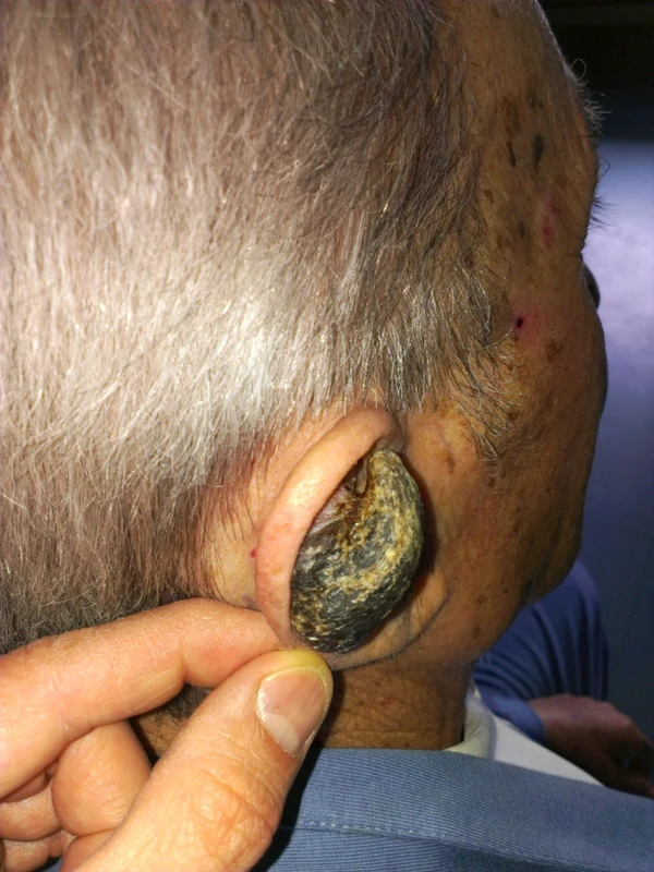
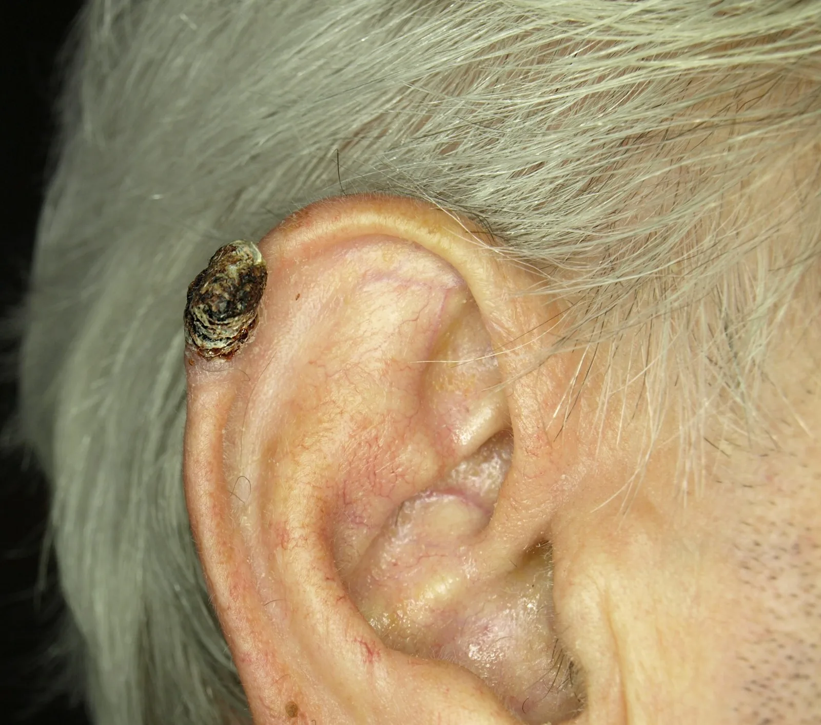
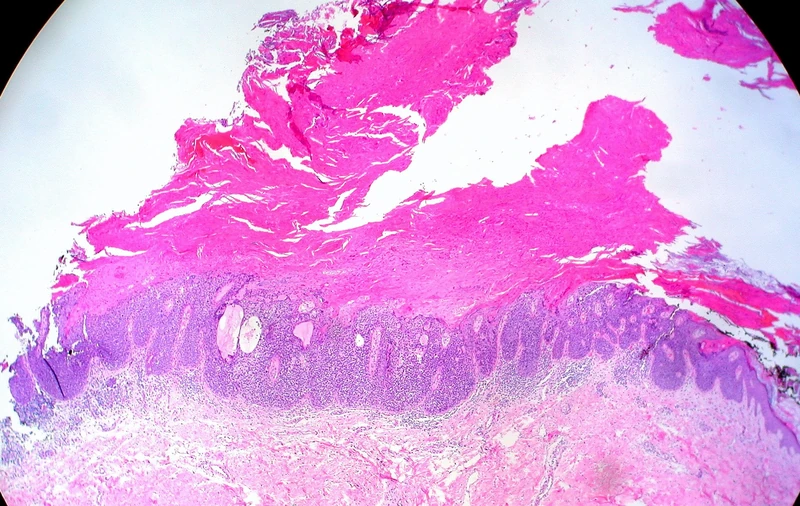 Histology
Definition
A cutaneous horn (cornu cutaneum) is a clinical description, not a diagnosis in its own right: a hard, conical projection of compact keratin resembling an animal's horn. What matters is the lesion at the base – it may be benign, premalignant or malignant.
Classification
By the lesion at the base:
- benign: seborrhoeic keratosis, viral wart, epidermal naevus, molluscum contagiosum, ostraceous psoriasis, hypertrophic lichen planus
- premalignant or malignant: actinic keratosis, Bowen's disease, keratoacanthoma, squamous cell carcinoma, rarely melanoma
Occurrence & epidemiology
Cutaneous horns occur mainly in older people (peak between 60 and 70 years) and more often in fair skin (phototypes I–II). Men and women are affected about equally often, but the base is more often malignant in men. In two histological series, the proportion of premalignant or malignant base lesions was 38.9% (643 horns) and 58.56% (222 horns).
Clinical features
- Straight or curved, hard, yellow-brown horn; the side is often terrace-like or oyster shell-like with horizontal ridges, and the base flat, raised or crater-like.
- Typically more than twice as tall as the width of its base; size from a few millimetres to several centimetres.
- Usually solitary, mainly on sun-exposed skin (head, ears, backs of the hands, forearms); usually asymptomatic.
- Clues to squamous cell carcinoma (none is conclusive): pain, large size, induration or redness of the base, a wide base or low height-to-base ratio, lack of terrace formation.
Histology
Compact hyperkeratosis, often with acanthosis. Orderly, horizontally parallel layers of keratin favour a benign lesion; erratic growth suggests a rapidly growing malignant lesion. The base shows the features of the underlying lesion.
Diagnosis
The horn itself is recognised clinically. Because benign and malignant base lesions cannot be reliably distinguished clinically, histological examination of the base of the horn is decisive.
Differential diagnoses
Keep learning in the app
References (selection)
Cross-references
Mentioned in
Note: This page is intended for medical education and does not replace diagnosis or treatment decisions in individual cases.