Blue naevus
- Synonyms
- Naevus caeruleus, blauer Naevus, Jadassohn-Tièche-Nävus, blue nevus, blue mole
- Specialty
- Dermatology · Skin tumours
- Images
- Clinical 1 · Histology 4
- In the app
- 1 flashcard
- Last updated
- 10/2026 · Dr. Pascal Bafteh
Contents
Images (5)
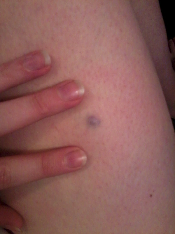
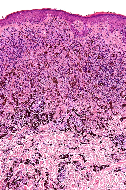 Histology
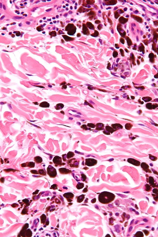 Histology
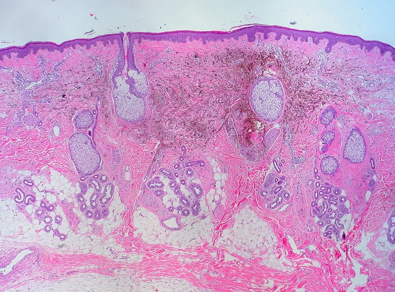 Histology
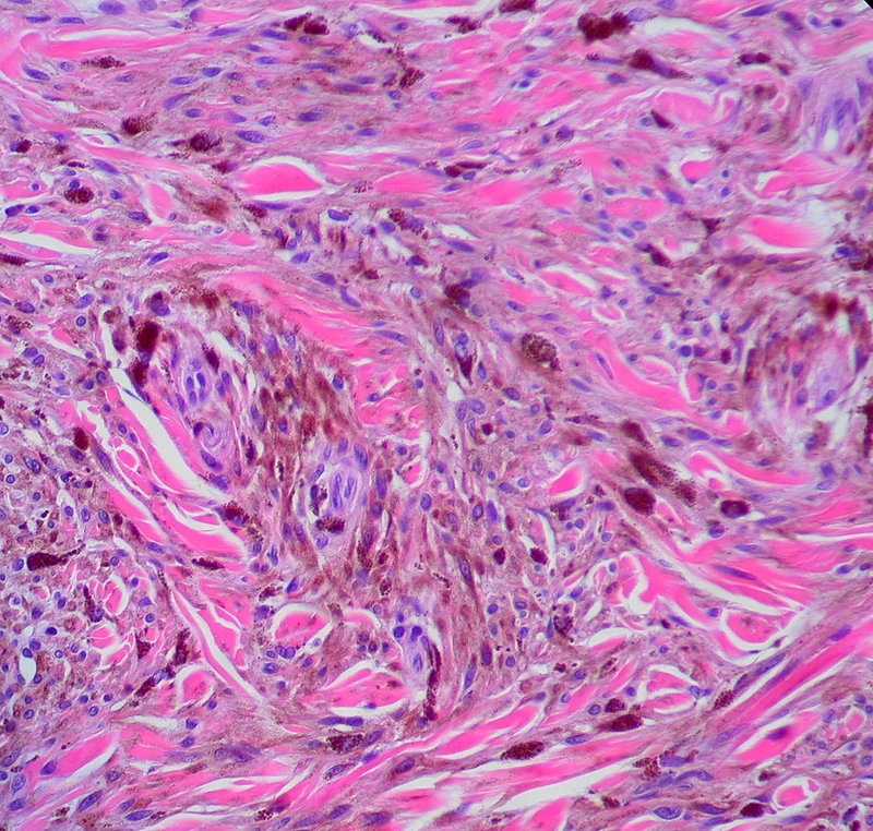 Histology
Definition
The blue naevus is a benign melanocytic tumour of pigmented, spindle-shaped melanocytes in the dermis. The blue colour is classically explained by light scattering from the deep-seated pigment (Tyndall effect); more recent work suggests rather a reduced scatter of red light.
Clinical features
- Common blue naevus: single, round to oval, blue to blue-grey, smooth macule, papule or plaque usually 0.5–1 cm in size, often on the back of the hand or foot, head or buttocks; usually appears in childhood or adolescence.
- Cellular blue naevus: more nodular and usually at least 1 cm in size.
- Common blue naevi remain unchanged throughout life; cellular blue naevi can very rarely become malignant (malignant blue naevus, a type of melanoma).
Diagnosis
- Dermoscopy: homogeneous, structureless steel-blue pigmentation.
- Distinction from nodular or metastatic melanoma, traumatic tattoo, vascular lesions (e.g. venous lake), dermatofibroma, Kaposi sarcoma and naevus of Ota/Ito; histological confirmation if the diagnosis is uncertain (spindle-shaped, heavily pigmented melanocytes in the mid-dermis, SOX10 and Melan-A positive).
Differential diagnoses
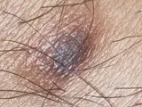
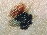
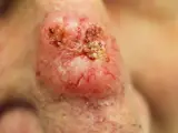
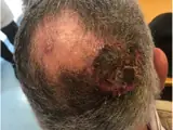
Image credits for the thumbnails
- Dermatofibroma: Jmarchn, “Pigmented and tenderness dermatofibroma”, CC BY-SA 3.0, via Wikimedia Commons · CC BY-SA 3.0 · Source
- Melanoma: National Cancer Institute (unknown photographer), “Melanoma (4)”, Public Domain, via Wikimedia Commons · Public domain · Source
- Basal cell carcinoma: Bin im Garten, “Basaliom am Nasenrücken 2”, CC BY-SA 3.0, via Wikimedia Commons · CC BY-SA 3.0 · Source
- Cutaneous angiosarcoma: Messias H, Martins M, Zagalo C et al. (Cancer Rep (Hoboken) 2022), CC BY 4.0, via PubMed Central (PMC9575495) · CC BY 4.0 · Source
Treatment principles
- The common blue naevus is benign and requires no treatment.
- Excision with histological examination is indicated for new, growing or nodular lesions and in cases of diagnostic uncertainty, particularly to exclude melanoma.
- For larger cellular blue naevi, complete excision is often recommended because differentiation is more difficult.
Specific treatment regimens with drugs and doses are covered in the DermaFuchs app.
Keep learning in the app
References (selection)
Cross-references
Keep learning: test yourself
One question from the DermaFuchs app on Blue naevus – tap an answer.
Question 1 of 1
Which gene mutation is associated with blue nevus?
Answer
Correct: GNAQ. A mutation in the GNAQ gene is commonly found in blue nevi, leading to their characteristic appearance.
More questions like these – with explanations and spaced repetition – in the app.
Note: This page is intended for medical education and does not replace diagnosis or treatment decisions in individual cases.