Lupus erythematosus gesicht
Etiopathogenesis
- Autoimmune disease with production of autoantibodies against nuclear material (dsDNA, Sm antigen, anti-SSA/SSB, ribonucleoproteins).
- Triggers: Classically, genetics + infections + UV + medications are involved, with EBV acting via molecular mimicry.
- UV mechanism: UV light hits cell nuclei → apoptosis with release of nuclear components → more "substrate" for autoantibodies → inflammation.
- Additionally relevant:
- Plasmacytoid DCs recognize DNA as DAMPs → IFN-α production
- Neutrophil extracellular traps (NETs) as a source of autoantigens
Classification of Cutaneous Lupus Erythematosus (CLE)
All forms with increasing risk for SLE.
Acute Cutaneous LE (ACLE)
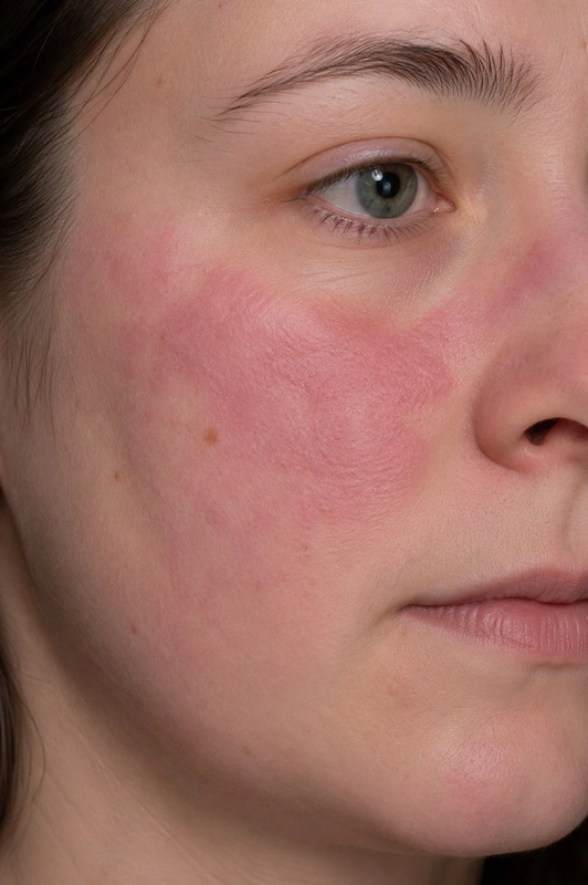
- Clinical features: Typical is a malar rash on the cheeks + nasal bridge, often with mucosal ulcers.
- Course: mostly part of an SLE (risk of systemic involvement ~ 80 %).
Subacute Cutaneous LE (SCLE)
- Patients: predominantly Caucasians, often with anti-Ro/SSA antibodies (~ 70–90 %).
- Clinical features: Typical are annular scaling plaques on the décolleté + shoulders, light-exposed areas, subacute.
- Risk of systemic involvement: approx. 15–50 % with often mild presentation.
Chronic Cutaneous LE (CCLE)
Discoid Lupus Erythematosus (DLE)
- Patients: more frequently dark-skinned patients.
- Clinical features: Typical are disc-shaped scarring plaques with central atrophy + adherent scaling and thimble phenomenon, often scarring alopecia on the scalp.
- Histopathology: The hallmark is interface dermatitis — vacuolar degeneration of the basal cell layer with apoptotic keratinocytes; in addition follicular plugging + thickened PAS-positive basement membrane + dermal mucin deposition and a perivascular and periadnexal lymphocytic infiltrate.
- Risk of systemic involvement: approx. 5 %.
Lupus Erythematosus Profundus (Lupus panniculitis)
- Clinical features: Typical are deep painful nodules with adipose tissue atrophy, frequently on cheeks, shoulders, hips.
Chilblain Lupus (Lupus pernio cutaneus)
- Clinical features: Typical are bluish-livid nodules on acral parts with cold triggering — cold- and moisture-dependent, tender papules and plaques on fingers, toes, tip of the nose and ears, ulceration possible, regression during the warm season.
- Classification: a distinct variant of chronic cutaneous LE (CCLE); overlap with DLE is common and progression to SLE is possible → monitor ANA/ENA, complete blood count and urinalysis.
Intermittent Cutaneous LE — Lupus Tumidus
- Clinical features: Typical are edematous non-scaling plaques with healing without scars — important differential diagnosis to PLD!
Transition & Follow-up
- Transition from SCLE / ACLE to SLE is not uncommon → therefore annual monitoring of CBC, urinalysis, and autoantibody profile for at least 5 years from the initial CLE diagnosis.
- Renal monitoring: For screening for renal involvement the urinalysis from a spot urine sample (dipstick, sediment, protein/creatinine ratio) is sufficient — a 24-hour urine collection is not required for this; it only becomes relevant for an abnormal screening result or to quantify established lupus nephritis.
- Anti-dsDNA: not only a diagnostic marker but an activity and follow-up parameter — rising titers together with falling complement (C3/C4) indicate a flare or the transition to SLE, in particular renal activity. Anti-Sm and anti-Ro/SSA, by contrast, remain largely titer-stable and are unsuitable for monitoring disease course.
- Hydroxychloroquine (HCQ): maximum 5 mg/kg actual body weight (DGRh / EULAR 2024 — previously 6.5 mg/kg ideal body weight); if no response after 3–6 months, additionally Quinacrine 100–200 mg/d.
- Ophthalmologic monitoring (HCQ retinopathy): baseline + annually after 5 years (OCT, visual acuity, visual field).
Differential diagnoses
- Atopic dermatitis (atopic eczema)
- Rosacea
- Actinic keratosis
- Discoid lupus erythematosus (CDLE)
- Seborrhoeic dermatitis
- Subacute Cutaneous Lupus Erythematosus (SCLE)
- Lupus Tumidus
- Dermatomyositis
Practise Lupus erythematosus gesicht in the app
Flashcards with spaced repetition, exam questions and spot-the-diagnosis on this topic – in DermaFuchs, free of charge.
In the DermaFuchs app: 4 flashcards · 3 clinical images
Open in browser Download on the App StoreSources (selection)
- AWMF S2k CLE 2023
- Aringer M, Costenbader K, Daikh D, et al. 2019 European League Against Rheumatism/American College of Rheumatology Classification Criteria for Systemic Lupus Erythematosus. Arthritis Rheumatol. 2019;71(9):1400-1412.
More connective tissue diseases
Note: This page is intended for medical education and does not replace diagnosis or treatment decisions in individual cases. Treatment and follow-up content is available in the app.