Favre-Racouchot disease
Exam relevance: in 1 of more than 50 board exam reports · rank 93
- Synonyms
- Favre–Racouchot syndrome
- Specialty
- Dermatology · Environmental & photodermatoses
- Images
- Clinical 1 · Dermoscopy 1 · Histology 1
- In the app
- 2 flashcards
- Last updated
- 10/2026 · Dr. Pascal Bafteh
Contents
Images (3)
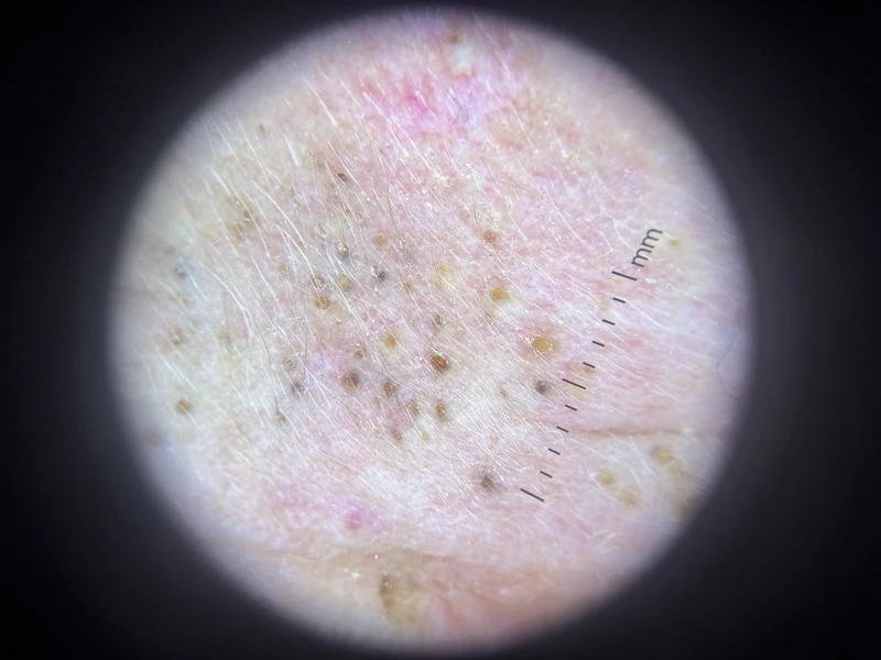 Dermoscopy
 Histology
Definition
Definition & Cause
- Favre-Racouchot disease (nodular cutaneous elastosis with cysts and comedones) = a consequence of chronic UV damage: solar elastosis with numerous open and closed comedones and small cysts in photodamaged facial skin.
- The decisive sentence: The black dots in Favre-Racouchot disease are solar comedones caused by chronic UV damage, not an expression of acne.
- Who is affected: older people with many years of cumulative sun exposure; men more often than women. Smoking is regarded as an additional contributing factor.
- Why "elastoidosis nodularis et cystica": the old name describes exactly what is seen — nodular-cystic remodelled, elastotic skin.
Clinical features
Clinical Presentation
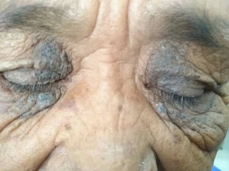
- Sites: Sites of predilection are periorbital, temple and zygomatic region, i.e. the light-exposed surfaces.
- Lesions: numerous open comedones ("blackheads"; the colour comes from melanin in the widened follicular ostium), plus closed comedones and small cysts.
- The background: yellowish thickened skin with coarse pores and a coarsened wrinkle pattern — this is the solar elastosis itself. Alongside it, solar lentigines from the same cause are regularly present.
- What is missing: Against acne speaks the complete absence of inflammatory papules and pustules.
- Symptoms: none. It does not itch and does not hurt — patients come because of the appearance, or the finding is noticed incidentally.
Why the Finding Matters
- Check as well: actinic keratoses, field cancerisation, basal cell carcinoma and squamous cell carcinoma — the comedones themselves are harmless, their neighbourhood is not.
- An opening for the conversation: consistent sun protection and smoking cessation are easy to argue for on the basis of this visible finding.
Histology
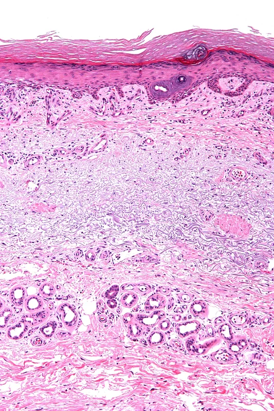
- Pronounced solar elastosis of the upper and mid dermis, together with markedly dilated follicular infundibula filled with lamellar keratin; cystically dilated follicular segments. Signs of inflammation are typically absent.
Exam essentials
- Key point: Favre–Racouchot disease = solar elastosis plus large open comedones and cysts on the temples and lateral periorbital area – typically in older men with long-term UV exposure, often smokers.
- Classic picture: yellowish, thickened skin lateral to the eyes with symmetrical black comedones, usually without inflammatory papules or pustules.
- Histology: marked actinic (solar) elastosis of the dermis, dilated follicular openings with keratin plugs, little inflammation.
- Distinction from acne vulgaris: older age, temporal/periorbital location, absence of inflammation and the background of chronic sun damage.
Differential diagnoses
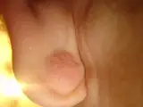
 Sebaceous hyperplasia
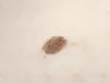
 Epidermoid cyst
 Seborrhoeic keratosis
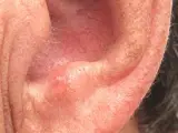 Actinic keratosis
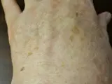 Solar lentigo (age spot)
 Milia (milium)
Image credits for the thumbnails
- Sebaceous hyperplasia: Klaus D. Peter, Wiehl, “Seborrhoeic dermatitis2” (Commons label differs from the diagnosis), CC BY 3.0 DE, via Wikimedia Commons · CC BY 3.0 DE · Source
- Epidermoid cyst: unknown (Commons user), “Epidermal Cyst ear”, CC BY-SA 3.0, via Wikimedia Commons · CC BY-SA 3.0 · Source
- Seborrhoeic keratosis: Assafn, “Seborrheic keratosis closup”, CC BY-SA 4.0, via Wikimedia Commons · CC BY-SA 4.0 · Source
- Actinic keratosis: Future FamDoc, “Actinic keratosis on ear”, CC BY-SA 4.0, via Wikimedia Commons · CC BY-SA 4.0 · Source
- Solar lentigo (age spot): Beyond My Ken, “Liver spots, light-skinned Caucasian man, age 63”, CC BY-SA 4.0, via Wikimedia Commons · CC BY-SA 4.0 · Source
- Milia (milium): Serephine, “Newborn Milia (Milk Spots)”, CC0, via Wikimedia Commons · CC0 · Source
Related conditions: how to tell them apart
- Actinic keratosis – another sign of chronic sun damage – examine the same skin areas
- Solar lentigo (age spot) – sun damage with pigmentation instead of comedones
- Sebaceous hyperplasia – yellowish papules with a central dell, no open comedones
- Acne vulgaris – comedones with inflammatory lesions, younger age
- Cutaneous squamous cell carcinoma – on sun-damaged skin, consider indurated or growing nodules
Treatment principles
- The basis is consistent UV protection and smoking cessation; these prevent progression but do not reverse existing changes.
- Topical retinoids, which loosen the keratin plugs, are first line; comedones and small cysts are expressed mechanically or curetted.
- Marked cases may be treated with ablative procedures such as dermabrasion or ablative laser; antibiotic acne therapy is of no use, as there is no inflammation.
Specific treatment regimens with drugs and doses are covered in the DermaFuchs app.
Keep learning in the app
References (selection)
Cross-references
Keep learning
Keep learning with flashcards, exam questions and spot diagnoses in the DermaFuchs app – free, in your browser or as an app.
Related pages: Acne comedonica · Sebaceous hyperplasia · Epidermoid cyst
Note: This page is intended for medical education and does not replace diagnosis or treatment decisions in individual cases.