Cutaneous tuberculosis
- Synonyms
- Tuberculosis cutis, skin tuberculosis
- Specialty
- Dermatology · Inflammatory skin diseases
- Images
- Clinical 2
- In the app
- 3 flashcards
- Last updated
- 10/2026 · Dr. Pascal Bafteh
Contents
Images (2)
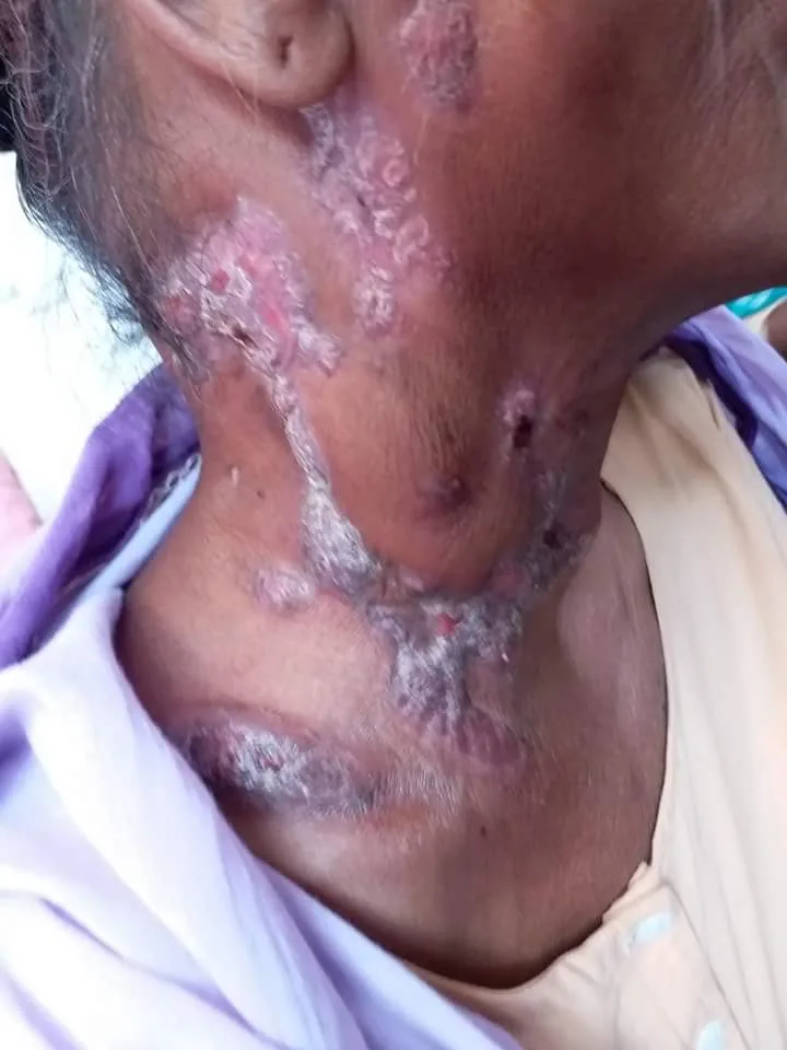
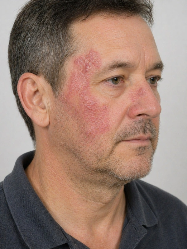 AI illustration
Definition
Cutaneous tuberculosis is an infection of the skin with Mycobacterium tuberculosis, rarely M. bovis. Its clinical picture depends on the route of infection (direct inoculation from outside, extension from an underlying focus or haematogenous spread) and on host immunity. In addition there are tuberculids – hypersensitivity reactions that typically develop in patients with moderate to high immunity to the tubercle bacillus.
- Lupus vulgaris (post-primary, slowly progressive with lupus nodules, diascopy: "apple jelly" phenomenon)
- Scrofuloderma (cutaneous manifestation of lymph node/bone TB)
- Tuberculosis cutis verrucosa (exogenous infection, hyperkeratotic plaques)
- Erythema induratum Bazin (vasculitis-like TB reaction on calves)
Diagnosis
- Biopsy with histology (tuberculoid granulomas with or without caseation), Ziehl-Neelsen stain, PCR and tissue culture.
- Interferon-gamma release assay or tuberculin skin test, chest X-ray, search for organ tuberculosis.
Differential diagnoses
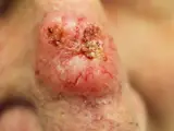
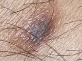
Image credits for the thumbnails
- Granuloma annulare: Kreuter et al., “Generalized granuloma annulare”, CC BY-SA 2.0, via Wikimedia Commons · CC BY-SA 2.0 · Source
- Actinic keratosis: Future FamDoc, “Actinic keratosis on ear”, CC BY-SA 4.0, via Wikimedia Commons · CC BY-SA 4.0 · Source
- Basal cell carcinoma: Bin im Garten, “Basaliom am Nasenrücken 2”, CC BY-SA 3.0, via Wikimedia Commons · CC BY-SA 3.0 · Source
- Dermatofibroma: Jmarchn, “Pigmented and tenderness dermatofibroma”, CC BY-SA 3.0, via Wikimedia Commons · CC BY-SA 3.0 · Source
Treatment principles
- Cutaneous tuberculosis is treated like organ tuberculosis with antituberculous combination therapy over several months, ideally after microbiological confirmation with susceptibility testing.
- As tuberculosis of other organs is not uncommonly present at the same time, it is actively sought; treatment is multidisciplinary with infectious diseases or respiratory medicine.
- Tuberculids such as erythema induratum also respond to antituberculous therapy.
Specific treatment regimens with drugs and doses are covered in the DermaFuchs app.
Keep learning in the app
References (selection)
Cross-references
Keep learning
Keep learning with flashcards, exam questions and spot diagnoses in the DermaFuchs app – free, in your browser or as an app.
Related pages: Granuloma annulare · Actinic keratosis · Basal cell carcinoma
Note: This page is intended for medical education and does not replace diagnosis or treatment decisions in individual cases.