Porokeratosis
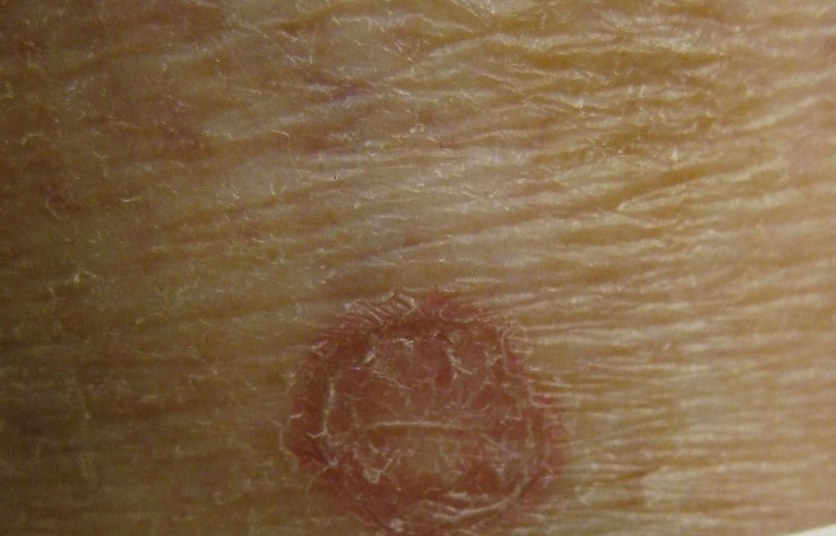
Porokeratosis — peripheral ridge and cornoid lamella (mini-section)
- Clinical features: sharply demarcated, centrally atrophic lesions with a ridge-like raised border — „like a wall"; this peripheral ridge is the clinical hallmark and appears under the lens as a narrow double furrow.
- Histology: cornoid lamella — an obliquely oriented compact column of parakeratosis over an area with loss of the granular layer; it is the exact histological correlate of the clinical border.
- Variants: porokeratosis of Mibelli (solitary, large, often in childhood), disseminated superficial actinic porokeratosis (DSAP) on sun-exposed extensor surfaces, porokeratosis palmaris et plantaris disseminata, linear porokeratosis.
- Caution: risk of degeneration to squamous cell carcinoma, especially in large, long-standing and linear lesions → follow-up, biopsy on growth, induration or erosion.
Differential diagnoses
- Actinic keratosis
- Psoriasis vulgaris (plaque psoriasis)
- Cornu cutaneum
- Verruca vulgaris
- Dermatofibroma
- Bowen's Disease
- Verruca vulgaris
- Granuloma annulare
Practise Porokeratosis in the app
Flashcards with spaced repetition, exam questions and spot-the-diagnosis on this topic – in DermaFuchs, free of charge.
In the DermaFuchs app: 3 flashcards · 6 clinical images
Open in browser Download on the App StoreMore skin tumours
- Basal cell carcinoma
- Melanoma
- Lentigo maligna
- Cutaneous squamous cell carcinoma
- Actinic keratosis
- Actinic cheilitis
- Merkel cell carcinoma
- Microcystic adnexal carcinoma
Note: This page is intended for medical education and does not replace diagnosis or treatment decisions in individual cases. Treatment and follow-up content is available in the app.