Mycosis fungoides
Exam relevance: in 6 of more than 50 board exam reports · rank 24
- Synonyms
- kutanes T-Zell-Lymphom, CTCL, cutaneous T-cell lymphoma
- Specialty
- Dermatology · Skin tumours
- Images
- Clinical 3 · Histology 1
- In the app
- 7 flashcards
- Last updated
- 10/2026 · Dr. Pascal Bafteh
Contents
Images (4)
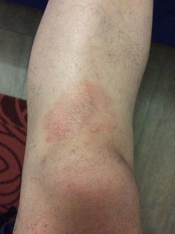
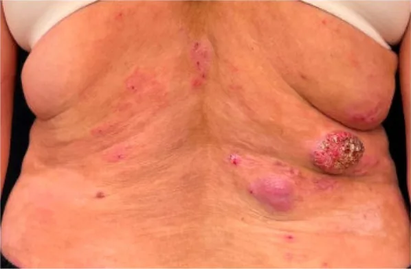
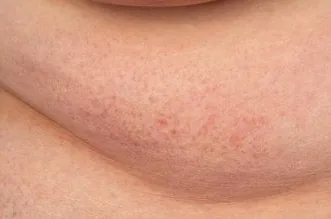
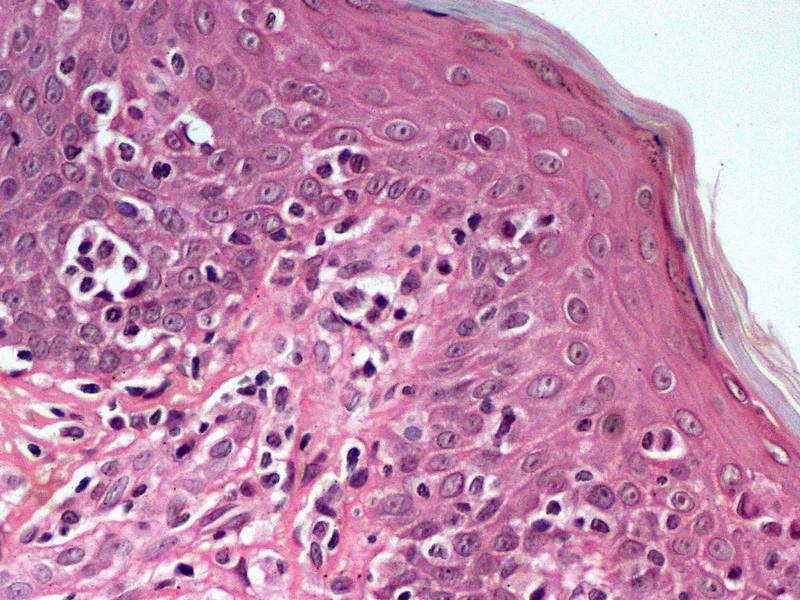 Histology
Definition
Definition & Epidemiology
- Mycosis fungoides (MF) = most common cutaneous T-cell lymphoma (CTCL), originating from CD4-positive helper memory T-cells with skin tropism.
- Sézary syndrome (SS) = leukemic variant with erythroderma + circulating atypical T-cells + generalized lymphadenopathy.
- Incidence of MF ~ 0.5 / 100,000 / year; mostly 50–60 years of age; M:F ≈ 2:1.
Clinical features
Clinical Presentation by Stage
- Patch: reddish-brown, non-infiltrated macules with fine lamellar scaling, often on buttocks / trunk
- Plaque: infiltrated erythematous plaques, sharply demarcated
- Stage IA / IB is defined not by patch versus plaque but by the body surface area involved: IA < 10 % BSA (T1), IB ≥ 10 % BSA (T2)
- Tumor (IIB): exophytic nodules / tumors, often ulcerated
- Erythroderma (III): > 80 % BSA, pruritus, "L'homme rouge"
- Sézary (IV): + atypical circulating cells, lymphadenopathy
Sézary Syndrome
- Triad: Erythroderma + generalized lymphadenopathy + atypical circulating Sézary cells.
- Excessive pruritus ("Sézary pruritus") — scratch-induced lichenification.
- Folliculotropic variant with alopecia + comedones + mucinosis.
Diagnosis
Diagnostics
Histology
- Characteristic but not obligatory correlate Pautrier microabscesses — intraepidermal CD4+ T-cell aggregates without significant spongiosis.
- Epidermotropism of atypical T-cells without spongiosis (DD to eczema).
- In advanced stages, dense band-like infiltrate in the upper dermis.
Immunohistochemistry + Clonality
- CD3+/CD4+/CD45RO+ T-helper cells, partial CD7 loss.
- TCR gene rearrangement PCR (T-cell clonality) — supports diagnosis, not solely diagnostic.
Flow Cytometry (peripheral blood)
- Sézary cell phenotype CD4+/CD7−/CD26− — atypical T-cells with CD7 and CD26 loss.
- Sézary diagnosis: ≥ 1000 Sézary cells/µl OR CD4:CD8 ratio > 10 OR ≥ 40 % CD4+/CD7 loss.
Tumor Burden Markers
- Tumor burden markers LDH + β2-microglobulin — as a surrogate for tumor mass.
- Staging: TNMB system (Olsen 2007), mSWAT score (BSA × lesion severity).
Differential diagnoses
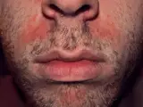 Seborrhoeic dermatitis
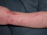 Atopic dermatitis (atopic eczema)
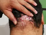 Folliculitis decalvans
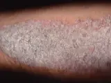 Psoriasis vulgaris (plaque psoriasis)
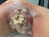 Primary cutaneous anaplastic large cell lymphoma (pcALCL)
- Primary cutaneous aggressive epidermotropic CD8-positive cytotoxic T-cell lymphoma
- Primary cutaneous diffuse large B-cell lymphoma (NOS)
- Erythroderma (exfoliative dermatitis)
Image credits for the thumbnails
- Seborrhoeic dermatitis: Roymishali, “Seborrhoeic dermatitis highres”, CC BY-SA 3.0, via Wikimedia Commons · CC BY-SA 3.0 · Source
- Atopic dermatitis (atopic eczema): Eisfelder, “Atopic dermatitis child”, CC BY-SA 3.0, via Wikimedia Commons · CC BY-SA 3.0 · Source
- Folliculitis decalvans: Mohammad2018, “Foliculitis declavane new photo to avoid wrong diagnosis”, CC BY-SA 4.0, via Wikimedia Commons · CC BY-SA 4.0 · Source
- Psoriasis vulgaris (plaque psoriasis): MediaJet, “An Arm Covered With Plaque Type Psoriasis”, CC BY-SA 3.0, via Wikimedia Commons · CC BY-SA 3.0 · Source
- Primary cutaneous anaplastic large cell lymphoma (pcALCL): Varela A, Sepúlveda S, Evans NE et al. (Cureus 2026), CC BY 4.0, via PubMed Central (PMC13061649) · CC BY 4.0 · Source
Treatment principles
- Treatment is stage-dependent and in most cases aims at disease control rather than cure.
- In early stages, skin-directed therapies predominate: topical glucocorticoids or cytostatics, phototherapy (narrowband UVB for patches, PUVA for plaques) and local radiotherapy.
- Advanced stages receive total skin electron beam therapy or systemic treatments such as retinoids, interferons, antibodies or chemotherapy, and extracorporeal photopheresis in Sézary syndrome.
- Planning is interdisciplinary; in selected patients, allogeneic stem cell transplantation can be curative.
Guideline: AWMF guideline register (no. 032-027)
Specific treatment regimens with drugs and doses are covered in the DermaFuchs app.
Keep learning in the app
References (selection)
Cross-references
Mentioned in
More skin tumours
Keep learning: test yourself
One question from the DermaFuchs app on Mycosis fungoides – tap an answer.
Question 1 of 1
What is the Sézary cell phenotype in Mycosis Fungoides and Sézary Syndrome?
Answer
Correct: CD4+/CD7−/CD26−. Sézary cells in Mycosis Fungoides and Sézary Syndrome exhibit a CD4+/CD7−/CD26− phenotype.
More questions like these – with explanations and spaced repetition – in the app.
Note: This page is intended for medical education and does not replace diagnosis or treatment decisions in individual cases.