Pilomatricoma (calcifying epithelioma of Malherbe)
- Synonyms
- Pilomatrikom, Epithelioma calcificans Malherbe, verkalkendes Epitheliom, Pilomatricoma, pilomatrixoma, calcifying epithelioma of Malherbe
- Specialty
- Dermatology · Skin tumours
- Images
- Clinical 1 · Histology 2
- In the app
- 1 flashcards
- Last updated
- 10/2026 · Dr. Pascal Bafteh
Contents
Images (3)
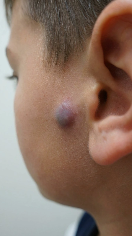 AI illustration
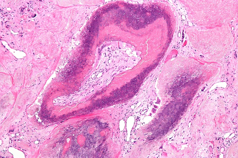 Histology
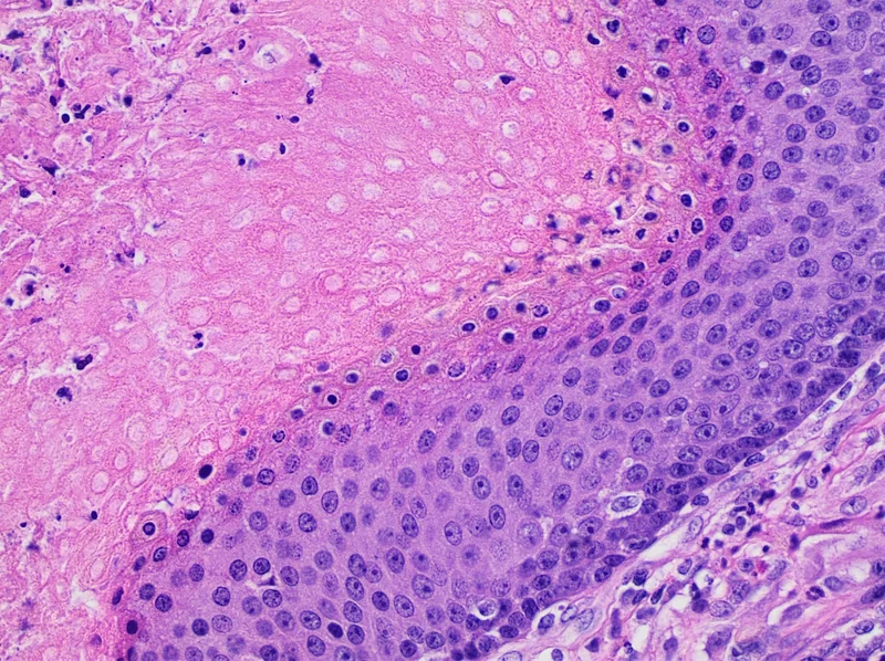 Histology
Definition
Pilomatricoma is a benign tumour arising from hair matrix cells. A characteristic feature is calcification, which makes the nodule feel hard.
Occurrence & epidemiology
It is most often diagnosed in children but also occurs in adults. In a series of 179 pilomatricomas of the head and neck, the mean age was 29.8 years and 45.3% of patients were younger than 18 years. Multiple pilomatricomas have been reported in association with myotonic dystrophy.
Aetiopathogenesis
The cause is a localised mutation in a hair matrix cell; most cases carry mutations in the CTNNB1 gene (beta-catenin), and BCL-2 is overexpressed.
Clinical features
- Solitary hard nodule, skin-coloured, whitish, reddish or purplish; usually 5–10 mm, stable for years or slowly growing to several centimetres.
- Mainly on the head and neck (series: neck 30.2%, cheek 16.8%, scalp 16.2%, brow and periorbital area 14.0%), but possible at any site.
- Usually asymptomatic, occasionally tender; calcification often gives an angulated shape ("tent sign").
- Rarely giant forms; pilomatrix carcinoma is very rare.
Histology
Well-circumscribed nodulocystic tumour of the lower dermis, not uncommonly extending into the subcutis. Basaloid cells resembling the hair matrix mature into anucleate eosinophilic shadow cells; calcification is frequent in these areas, with multinucleated giant cells at sites of rupture. A pigmented variant occurs in up to 11%.
Diagnosis
- Dermoscopy: central whitish or grey-blue structureless area, sometimes erythema and telangiectases.
- Ultrasound: detection of calcification (in 88.2% in a paediatric series); also visible on X-ray.
- Histological confirmation.
Differential diagnoses
Keep learning in the app
References (selection)
Cross-references
Note: This page is intended for medical education and does not replace diagnosis or treatment decisions in individual cases.