Primary cutaneous anaplastic large cell lymphoma (pcALCL)
- Synonyms
- pcALCL, PC-ALCL, kutanes ALCL, CD30-positives kutanes T-Zell-Lymphom, CD30-positive lymphoproliferative Erkrankung, cutaneous ALCL, CD30-positive cutaneous lymphoma
- Specialty
- Dermatology · Skin tumours
- Images
- Clinical 1 · Histology 2
- In the app
- 1 flashcards
- Last updated
- 10/2026 · Dr. Pascal Bafteh
Contents
Images (3)
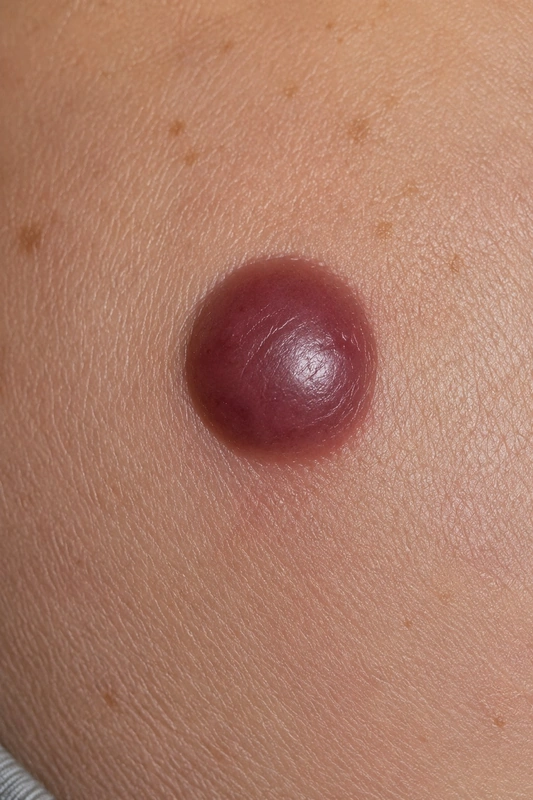 AI illustration
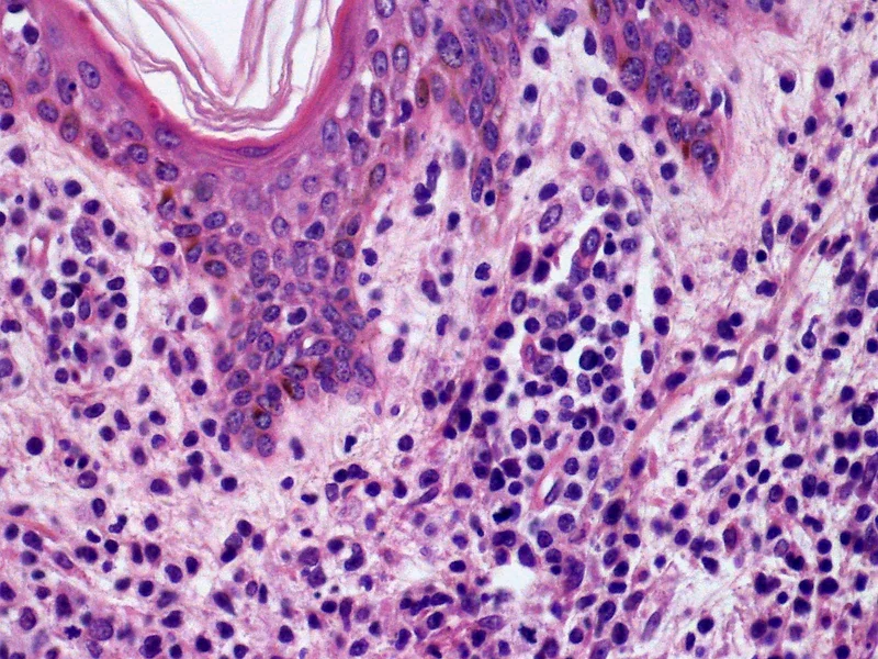 Histology
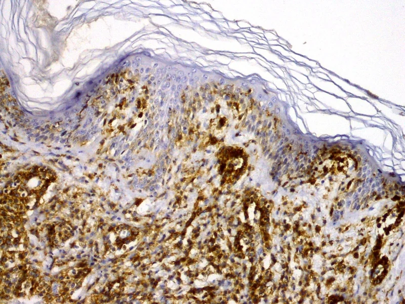 Histology
Definition
pcALCL is a rare, usually indolent cutaneous T-cell lymphoma composed of large, CD30-positive atypical lymphocytes. Together with lymphomatoid papulosis, it belongs to the primary cutaneous CD30-positive lymphoproliferative disorders and is the second most common cutaneous T-cell lymphoma after mycosis fungoides.
Occurrence & epidemiology
Men are affected about three times as often as women; most patients are 50–70 years old. Paediatric and congenital cases have been reported.
Aetiopathogenesis
The cause is unknown. About 30% of tumours carry a rearrangement of the DUSP22 gene (6p25.3).
Clinical features
- Usually a rapidly growing solitary nodule or plaque, red to violaceous, typically at least 2 cm in size and often ulcerated; usually asymptomatic.
- Multiple grouped or generalised nodules in about 20%.
- Any skin region including mucosal surfaces can be affected, most often the upper half of the body.
- No systemic symptoms (no fever, weight loss or night sweats).
- Partial or complete spontaneous regression in 20–40% after a median of 2 months; extracutaneous involvement, mostly of lymph nodes, in about 10%.
Histology
Diffuse dermal infiltrate of medium to large anaplastic or pleomorphic cells with abundant cytoplasm and horseshoe-shaped nuclei, with no or only focal epidermotropism, sometimes extending into the subcutis. At least 75% of tumour cells are CD30-positive; a clonal T-cell receptor gene rearrangement is found in 60–100%.
Diagnosis
- Skin biopsy with histology and immunohistochemistry.
- Dermoscopy: peripheral branching and polymorphous vessels on a pink-yellow structureless background.
- Staging (blood count, LDH, CT and/or PET-CT; TNM classification) to distinguish systemic ALCL with skin involvement.
- Lymphomatoid papulosis often cannot be distinguished on histology alone.
Differential diagnoses
Keep learning in the app
References (selection)
Cross-references
Note: This page is intended for medical education and does not replace diagnosis or treatment decisions in individual cases.